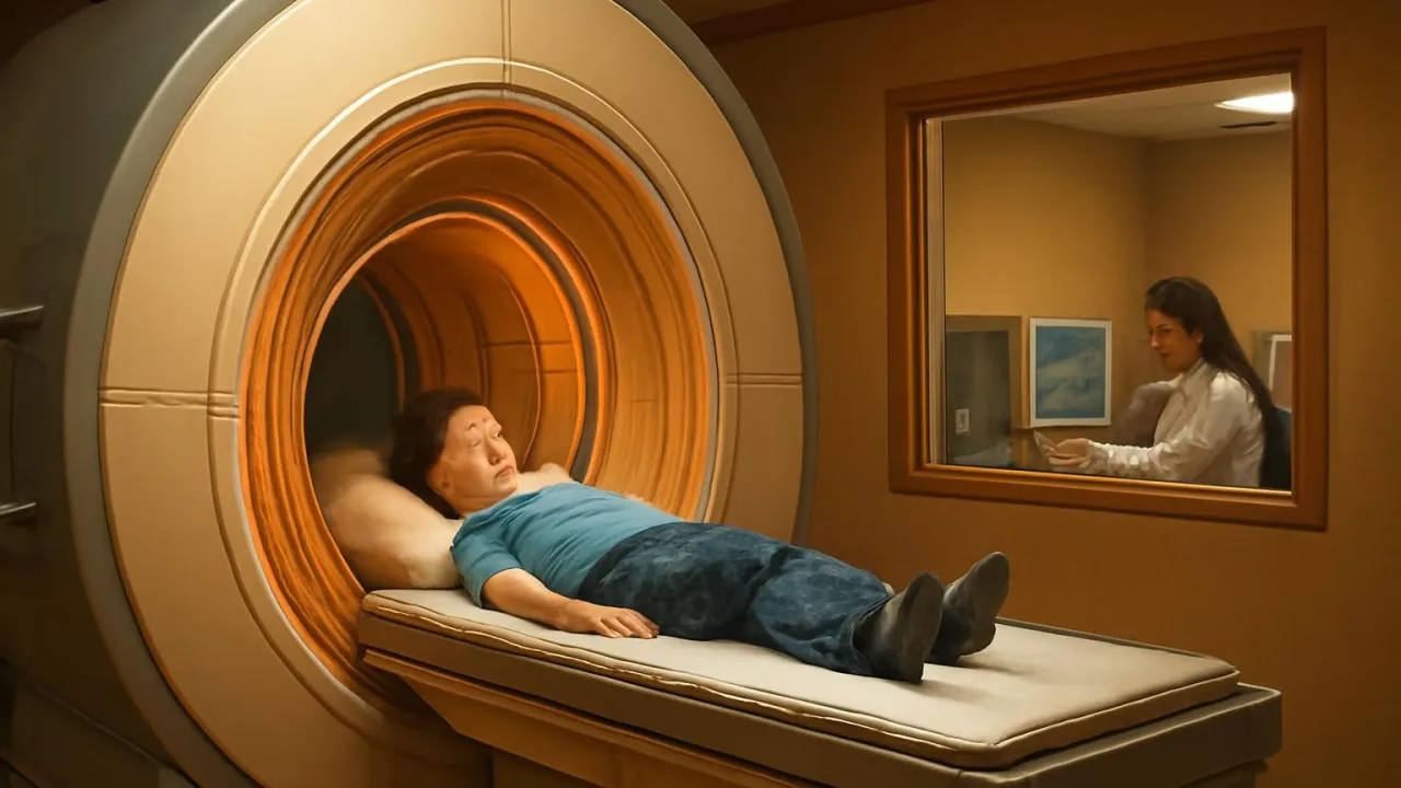
The MRI can even show your heart beating while you are inside!
✨ Word Sparks (Grade 2 Glossary)
Tap 🔊 to hear
radio waves
Invisible waves that carry energy
muscle
A soft part that helps you move
brain
The part that helps you think
🪝
Have you ever wondered how doctors see inside your body without opening it?
1
A Tunnel Full of Surprises
When you go inside an MRI, you hear thumping and knocking sounds. The machine is shaped like a big, round tunnel. You must lie very still so the pictures are clear.
2
Magnets and Pictures
The MRI uses strong magnets and to make pictures. These pictures show soft parts like your and . Doctors use them to find out what is wrong inside you.
3
Helping People Heal
MRI pictures help doctors plan how to help people get better. It does not hurt and does not use any x-rays. That makes it safe for kids and adults.
MRI scanners are so sensitive, they can see water moving inside your brain!
✨ Word Sparks (Grade 3 Glossary)
Tap 🔊 to hear
resonance
A special kind of vibration or echo
tissue
Soft parts that make up your body
diagnose
To find out what is wrong
🪝
What if a giant magnet could help doctors see your muscles and brain without a single cut?
1
Inside the MRI Tunnel
When you enter the MRI, you hear loud knocking and humming noises. The tunnel is a big cylinder that fits your whole body inside. You must stay very still so the machine can scan you. The MRI’s strong magnets create a safe but powerful field.
2
How the MRI Works
MRI stands for Magnetic Imaging, a breakthrough in medical technology. The scanner uses vibration and resonance from radio waves to make pictures of soft . Unlike x-rays, it does not use radiation, so it is safer for your body. Doctors can see details in your brain, muscles, and organs.
3
A World of Pictures
The MRI creates detailed images called scans that show what is happening inside. These scans help doctors find injuries, tumors, or other problems quickly. The machine can even show your heart beating or your brain thinking. MRI images are so clear, they can spot tiny changes in .
4
Changing Medicine Forever
The first full-body MRI scan was done in 1977 by Dr. Raymond Damadian. MRI technology changed how doctors and treat many illnesses. Now, millions of people around the world use MRI scans each year. This invention saves lives by helping doctors see what needs to be fixed inside the body.
2. Who helped create the first full-body MRI scan?
🤯 Mind-Blowing Fact • Grade 4
Superconducting magnets in MRI machines are cooled to nearly -452°F using liquid helium!
✨ Word Sparks (Grade 4 Glossary)
Tap 🔊 to hear
superconducting
A material that carries electricity with no resistance
hydrogen atom
The smallest part of water and your body
diagnosis
The process of finding out an illness
🪝
Imagine lying inside a giant humming tunnel, while invisible waves create a map of your insides—how does it work?
1
The Magnetic Tunnel Experience
When you lie inside an MRI scanner, you hear rhythmic thumps and buzzing as the machine works. The tunnel is a large, cylindrical chamber lined with powerful magnets. You must stay very still, because even tiny movements can blur the images. Technicians watch from a control room, making sure you are safe and comfortable throughout the scan.
2
The Science Behind MRI
MRI stands for Magnetic Resonance Imaging, a technology that uses physics to look inside the body. The scanner’s strong magnetic field lines up in your tissues, and radio waves make them vibrate. When the radio waves stop, the atoms send back signals, which the computer turns into detailed images. This process does not use any harmful radiation, making it safer than x-rays or CT scans.
3
A Revolution in Medicine
The first human MRI scan was performed in 1977 by Dr. Raymond Damadian and his team. Before MRI, doctors could not see soft tissues like the brain or muscles very clearly. Now, MRI images help diagnose injuries, tumors, and diseases quickly and accurately. Millions of MRI scans are performed worldwide each year, saving lives and improving treatments.
4
Global Impact and Future Possibilities
MRI technology has changed healthcare all over the world, making early possible for many illnesses. Scientists are now developing even faster MRI machines that can show how the brain works in real time. Hospitals, research centers, and sports teams all use MRI for different reasons. In the future, MRI may become even more advanced, helping people stay healthy in new ways.
1. Why do MRI machines use superconducting magnets?
2. What does MRI help doctors see best?
🤯 Mind-Blowing Fact • Grade 5
The strongest MRI scanners can create magnetic fields over 140,000 times stronger than a refrigerator magnet!
✨ Word Sparks (Grade 5 Glossary)
Tap 🔊 to hear
superconducting magnet
A super-strong magnet with no energy loss
resonance
When something vibrates at a special frequency
Tesla (T)
A unit that measures magnetic field strength
🪝
What if a machine could reveal the tiniest secrets inside your body—using only magnets and invisible waves?
1
Inside the MRI: A Sensory Adventure
As you slide into the cylindrical MRI scanner, you are surrounded by a powerful magnetic field up to 60,000 times stronger than Earth’s. The machine produces rhythmic thumping, clicking, and buzzing sounds as electric currents flow through tightly wound superconducting coils. Patients must remain perfectly still, sometimes for over an hour, to prevent image blurring from even the smallest movement. Technicians monitor the procedure from a shielded control room, communicating with you through speakers and microphones. The experience can feel futuristic, but every sound and sensation is part of a precise scientific process.
2
The Physics of Magnetic Resonance Imaging
MRI works by aligning the hydrogen nuclei in your body’s water molecules using a strong magnetic field. When a radiofrequency pulse is applied, these nuclei absorb energy and enter a higher energy state, a process called . As the pulse stops, the nuclei release energy, emitting radio signals that are detected by receiver coils. Sophisticated computers analyze these signals, constructing high-resolution, cross-sectional images of organs, muscles, and even the brain’s intricate structures. Unlike x-rays or CT scans, MRI uses no ionizing radiation, making it a safer choice for repeated imaging.
3
From Invention to Medical Revolution
Dr. Raymond Damadian, an American physician and scientist, built the first full-body MRI scanner in 1977 after discovering that cancerous tissue responded differently to magnetic fields. His prototype, called 'Indomitable,' paved the way for rapid advances in medical imaging. By the 1980s, MRI machines were being installed in hospitals worldwide, transforming how doctors diagnose strokes, tumors, joint injuries, and more. Today, over 100 million MRI scans are performed globally each year, saving countless lives through early detection and precise treatment planning.
4
Engineering Marvels and Quantitative Power
Modern MRI scanners use cooled with liquid helium to nearly absolute zero, eliminating electrical resistance and enabling fields of 1.5 to 7 Tesla (the unit of magnetic strength). Gradient coils change the magnetic field in tiny steps, allowing the machine to target specific body regions and create three-dimensional images. Advanced MRI techniques, like functional MRI (fMRI), can even map brain activity by tracking changes in blood flow, opening new frontiers in neuroscience and psychology. The engineering behind MRI is a blend of physics, electronics, and medical science.
5
The Future: Faster, Sharper, and More Accessible
Researchers are developing portable and ultra-high-field MRI systems that could make scanning quicker and available in more places, including remote clinics. Artificial intelligence is being used to interpret MRI data faster and with greater accuracy, reducing wait times and improving diagnoses. As MRI technology evolves, it continues to inspire new discoveries in biology, medicine, and engineering. The MRI scanner is not just a machine—it is a window into the living mysteries of the human body, forever changing the way we see ourselves.