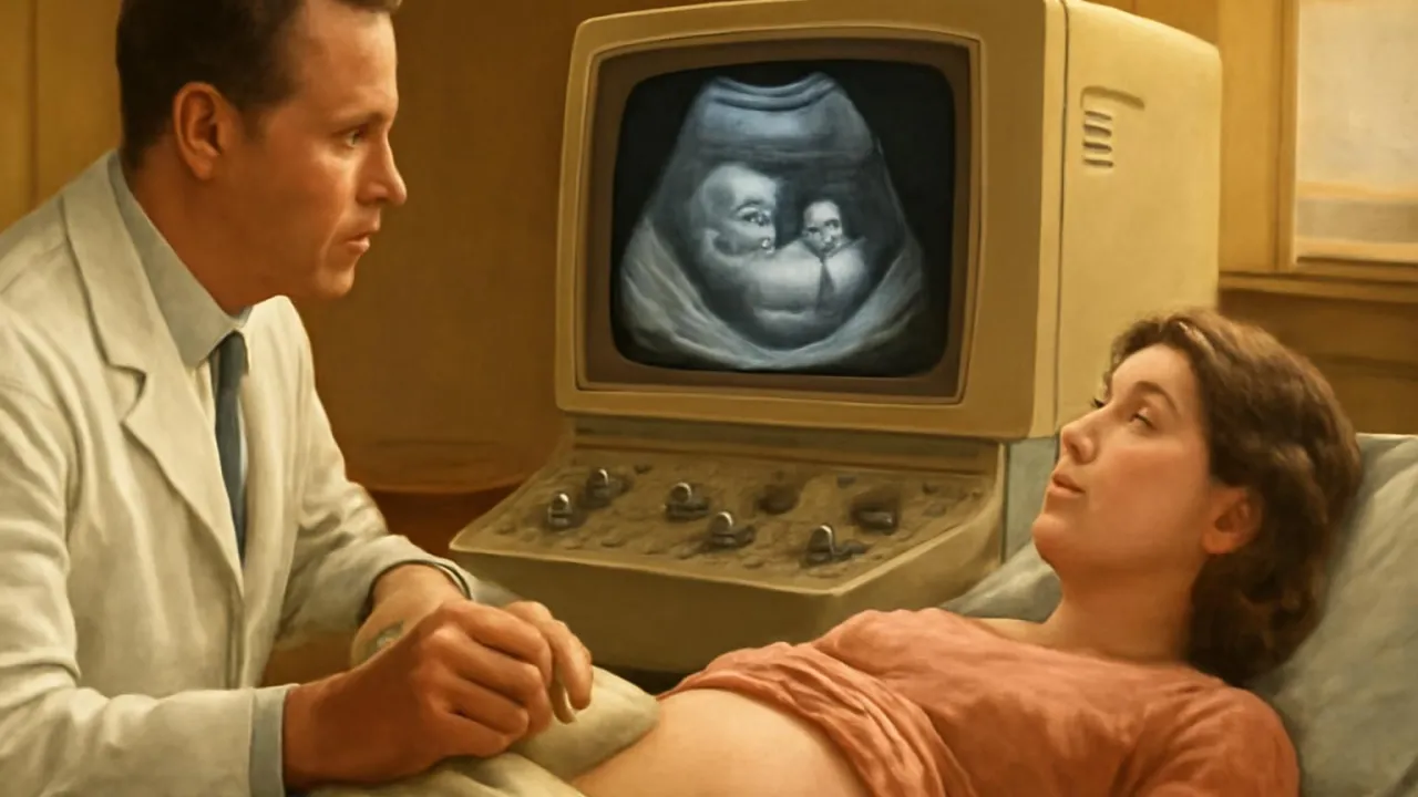
Ultrasound can show moving blood inside your veins and arteries!
✨ Word Sparks (Grade 2 Glossary)
Tap 🔊 to hear
echo
A sound that bounces back
organ
A special part inside your body
ultrasound
A way to see with sound waves
🪝
Have you ever heard a doctor make pictures with sound instead of light?
1
Sound Waves Make Pictures
A doctor slides a small probe on your skin, and the machine buzzes quietly. The probe sends sound waves into your body, and these waves bounce back when they hit something inside. The machine uses the to make a picture on the screen. You don’t feel the sound, but you can see the picture appear.
2
Why Is Ultrasound Special?
lets doctors see inside you without hurting or opening your body. They can watch a baby move or check your heart beating. It is safe, quick, and does not use any x-rays. Many people get ultrasounds when they visit the doctor.
3
What Do Doctors Look For?
Doctors use to check if babies are growing well or if look healthy. They can see hearts, livers, and even blood moving. This helps doctors know what care you need.
1. What does the ultrasound probe send into your body?
2. Why do doctors like using ultrasound?
🤯 Mind-Blowing Fact • Grade 3
The first ultrasound image of a baby was made in 1958 in Scotland!
✨ Word Sparks (Grade 3 Glossary)
Tap 🔊 to hear
vibration
A quick back-and-forth movement
sonogram
A picture made by ultrasound
non-invasive
Does not cut or hurt skin
🪝
What if you could see your heartbeat without even making a cut?
1
A Picture from Sound
Ultrasound imaging uses high-frequency sound waves to create pictures of the inside of your body. A doctor moves a device called a probe over your skin, which sends out these sound waves. When the waves hit something inside, like your heart or a baby, they bounce back as echoes. The machine turns these echoes into a moving image on a screen.
2
How Does Ultrasound Work?
The probe produces much faster than humans can hear, called ultrasound. These vibrations travel through soft tissue and bounce off denser parts, like organs or bones. The returning echoes carry information about how far away and how dense things are. The computer uses this data to build a real-time image, called a .
3
Why Is It Important?
Ultrasound is , which means it does not hurt or break the skin. Doctors use it to check on unborn babies, examine hearts, and find problems in organs. It is safer than x-rays because it uses sound instead of radiation. Many hospitals and clinics use ultrasound every day for quick and gentle checks.
4
A World of Uses
Besides medicine, ultrasound helps scientists study whales and dolphins by tracking their sounds underwater. Engineers use it to find cracks in metal pipes and airplane wings. This invention helps people stay healthy and keeps machines working safely.
The sound waves in medical ultrasound vibrate at over 1 million times per second!
✨ Word Sparks (Grade 4 Glossary)
Tap 🔊 to hear
acoustic
Related to sound or hearing
prototype
The first working model of something
echo
A sound that bounces back
🪝
Imagine a gentle humming probe revealing the hidden world beneath your skin—how does it work?
1
The Science of Sound Imaging
Ultrasound imaging relies on high-frequency sound waves, far above what humans can hear, to create pictures of the inside of the body. When a doctor places the probe on your skin, it sends rapid vibrations that travel through soft tissues and reflect off denser objects, such as organs or bones. These reflected sound waves, called , are collected by the probe and sent to a computer. The computer processes the echoes and forms a real-time image, known as a sonogram, on the screen.
2
A Medical Revolution
Before ultrasound, doctors had to use x-rays or surgery to see inside the body, which could be risky. The invention of medical ultrasound in the 1950s by Dr. Ian Donald and Tom Brown in Scotland changed everything. Their early used principles similar to sonar, which detects objects underwater. Ultrasound quickly became popular because it is non-invasive, painless, and does not use harmful radiation.
3
How Ultrasound Changed Medicine
Ultrasound is now essential for monitoring pregnancies, diagnosing heart conditions, and checking organs for disease. It lets doctors see moving blood, beating hearts, and growing babies—all without making a single cut. The technology is also used in emergency rooms, sports medicine, and even to guide doctors during surgery. Its safety and speed make it a favorite tool in hospitals worldwide.
4
Beyond the Hospital
Ultrasound imaging has applications far beyond medicine. Marine biologists use it to study whales and dolphins by tracking their sound signals. Engineers rely on ultrasound to inspect bridges and airplanes for hidden cracks or damage. This remarkable invention continues to help people and protect technology around the globe.
1. What makes ultrasound safer than x-rays for patients?
2. Who invented medical ultrasound imaging in the 1950s?
🤯 Mind-Blowing Fact • Grade 5
Doppler ultrasound can measure blood flow in vessels as small as 1 millimeter wide!
✨ Word Sparks (Grade 5 Glossary)
Tap 🔊 to hear
acoustic impedance
How much a material slows sound waves
Doppler effect
Change in sound pitch when things move
elastography
A scan that shows tissue stiffness
🪝
What if you could map the living body’s secrets using nothing but invisible sound waves?
1
The Physics Behind Ultrasound Imaging
Ultrasound imaging operates by transmitting high-frequency sound waves—typically between 2 and 18 megahertz—into the body using a transducer probe coated with conductive gel. These waves propagate through tissues at different speeds depending on their density and , reflecting back as echoes when they encounter boundaries between materials like muscle, fat, or bone. The probe detects these echoes, and a sophisticated computer algorithm reconstructs them into a two-dimensional grayscale image, known as a sonogram. Because the frequencies used are far above human hearing, patients experience no sensation from the sound itself.
2
Engineering a Medical Breakthrough
The first practical medical ultrasound device was developed in 1958 by Dr. Ian Donald and engineer Tom Brown in Glasgow, Scotland, adapting sonar technology originally designed to detect submarines. Their prototype used a water bath and mechanical arm to scan tissue samples, but modern machines use digital electronics and advanced signal processing. Today’s portable ultrasound units are compact, battery-powered, and capable of producing real-time three-dimensional images, revolutionizing medical diagnostics worldwide.
3
Clinical Impact and Global Reach
Ultrasound has become a cornerstone of modern medicine, enabling non-invasive monitoring of fetal development, cardiac function, and organ health. Its safety—since it uses no ionizing radiation—makes it ideal for repeated use, especially in prenatal care. Doppler ultrasound, a specialized form, can even measure the speed and direction of blood flow, aiding in the diagnosis of heart defects and vascular diseases. The World Health Organization considers ultrasound essential for basic healthcare in both developed and developing countries.
4
Beyond Human Health: Broader Applications
Ultrasound technology extends far beyond hospitals. Marine biologists use it to track whales and dolphins, while engineers employ ultrasonic testing to detect microscopic cracks in aircraft wings and bridges. In industry, ultrasonic cleaning uses high-frequency vibrations to remove dirt from delicate objects. The same principles even help archaeologists study ancient artifacts without damaging them, demonstrating ultrasound’s versatility across science and technology.
5
The Future of Ultrasound Innovation
Recent advances include portable handheld devices, artificial intelligence-assisted image analysis, and , which measures tissue stiffness to detect tumors. Researchers are developing high-intensity focused ultrasound (HIFU) for non-invasive treatments, such as destroying cancer cells or breaking up kidney stones. As technology progresses, ultrasound imaging will continue to provide clearer, faster, and safer ways to explore and heal the living world.