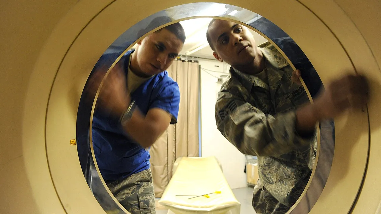
1. What shape does the outside of a hospital CT scanner look like?
2. What puts all the spinning X-ray pictures together for doctors?
🤯 Mind-Blowing Fact • Grade 2
Museum scientists also use CT scanners to peek inside wrapped Egyptian mummies and dinosaur fossils without breaking them open!
✨ Word Sparks (Grade 2 Glossary)
Tap 🔊 to hear
sensor
A part that detects light, heat, or sound
slice
One thin flat layer of a whole object
model
A detailed copy shown on a computer screen
🪝
Why couldn't ordinary flat X-rays show doctors everything hiding inside the human body?
1
More Than a Flat Shadow
Imagine shining a flashlight at your hand to make a flat shadow on the wall. Ordinary X-rays work like that shadow, so ribs in front can hide softer parts right behind them.
2
Spinning Around in a Circle
In 1971, British engineer Godfrey Hounsfield built a new kind of machine shaped like a giant doughnut. As a patient rests on a sliding table, a hidden X-ray tube spins smoothly in a full circle.
3
Stacking Picture Slices
catch beams from hundreds of angles, and a computer turns them into thin picture . Doctors can stack those slices on a screen to build a complete 3D without surgery!
1. What problem do ordinary flat X-ray pictures have?
2. Besides doctors, who else uses CT scanners to study fragile objects?
🤯 Mind-Blowing Fact • Grade 3
Engineers assign every tiny cube in a CT scan a brightness score on the Hounsfield scale, named directly after inventor Godfrey Hounsfield!
✨ Word Sparks (Grade 3 Glossary)
Tap 🔊 to hear
tomography
Making images of thin slices inside an object
gantry
The ring frame holding spinning scanner parts
prototype
The very first working test model built
🪝
What if you could inspect every single seed inside a watermelon without ever using a knife?
1
Seeing From Every Angle
Step into a hospital imaging room and you will spot a sleek ring called a . When the patient table glides through that opening, a hidden X-ray source and opposing detectors whirl silently in a 360-degree circle.
2
Two Brilliant Minds
Physicist Allan Cormack worked out the complex math needed to map internal tissues from outside beams. Soon after, British engineer Godfrey Hounsfield built the first working , scanning a patient's brain in London in October 1971.
3
What Tomography Means
The letters CT stand for computed , and the Greek root tomos means slice. Dense bone blocks more X-rays than soft muscle or air-filled lungs, creating measurable differences at every angle.
4
Fast Life-Saving Pictures
Powerful software solves thousands of equations to assemble cross-sectional images in moments. Emergency doctors rely on these crisp 3D views to spot tiny fractures, internal injuries, or blocked blood vessels.
1. What does the Greek root word 'tomos' in tomography mean?
2. Why do bones and lungs look different inside a CT scan?
🤯 Mind-Blowing Fact • Grade 4
Hounsfield's very first laboratory test machine took nine whole days to scan a preserved specimen and more than two hours for a computer to calculate one blurry image!
✨ Word Sparks (Grade 4 Glossary)
Tap 🔊 to hear
radiograph
An image produced on a surface by X-rays
helical
Shaped like a smooth corkscrew or spiral
voxel
A tiny three-dimensional cube in digital images
🪝
How did clever mathematics turn overlapping X-ray shadows into crystal-clear 3D maps of the human brain?
1
Overcoming the Shadow Problem
For seventy-five years after Wilhelm Roentgen discovered X-rays in 1895, doctors faced a frustrating limitation: conventional squashed a three-dimensional human body onto one flat photographic film. Inside the head, thick skull bone cast a bright white shadow that completely masked delicate soft brain tissue, forcing surgeons to guess where hidden injuries lay.
2
Math Meets Engineering
Working independently on two continents, physicist Allan Cormack developed mathematical formulas showing how X-ray absorption measured from many outside angles could reconstruct an interior map. Meanwhile in England, electrical engineer Godfrey Hounsfield paired a narrow X-ray beam with crystal detectors and a minicomputer to scan a patient in 1971—winning both pioneers the 1979 Nobel Prize.
3
Helical Spirals in Motion
Early hospital scanners rotated slowly step by step, pausing to unwind power cables after every single slice. Modern CT scanners use brushless slip-ring connectors inside the circular gantry so the heavy X-ray tube spins continuously several times per second while the motorized table glides smoothly forward, tracing an unbroken spiral path along the body.
4
Beyond Hospital Walls
High-speed computer algorithms translate millions of detector readings into three-dimensional volume pixels called that doctors can rotate or slice on screen from any direction. Today, besides guiding life-saving emergency trauma care and rapid heart exams, specialized CT scanners inspect jet turbine blades for microscopic cracks and reveal fragile dinosaur embryos still curled inside fossilized stone eggs.
1. Why couldn't standard flat X-rays clearly show soft brain tissue?
2. What allows a modern helical CT scanner to spin continuously without tangling its wires?
🤯 Mind-Blowing Fact • Grade 5
Spinning inside a modern high-speed CT scanner generates over 30 Gs of outward force—more than three times the G-force experienced by fighter pilots in sharp turns!
✨ Word Sparks (Grade 5 Glossary)
Tap 🔊 to hear
attenuation
Gradual weakening of a beam passing through matter
centrifugal
Pulling outward away from a spinning center
nondestructive
Testing an object without harming or breaking it
🪝
How do inverse mathematical transforms convert millions of invisible X-ray measurements into a beating human heart in less than a second?
1
Inside the Whirring Gantry
Behind the smooth composite cover of a hospital CT gantry, a two-ton rotating frame carries a high-voltage X-ray tube and an opposing curved arc of solid-state scintillation detectors. Driven by brushless direct-drive motors and slip-ring data links, the assembly completes nearly four revolutions per second—withstanding outward forces exceeding thirty times Earth's gravity without vibrating out of millimeter alignment.
2
Attenuation and the Radon Transform
As collimated fan-shaped X-ray beams traverse the body, each internal structure weakens—or attenuates—the photon stream according to its atomic number and electron density. Building independently upon Austrian mathematician Johann Radon's 1917 integral theorems, physicist Allan Cormack and engineer Godfrey Hounsfield demonstrated that sampling thousands of angular projections across 360 degrees allows exact cross-sectional reconstruction.
3
The Hounsfield Scale and Voxel Grids
Reconstruction algorithms—from classic filtered back-projection to modern AI-assisted iterative modeling—partition each sub-millimeter slice into a fine matrix of three-dimensional voxels. Every voxel receives a quantitative radiodensity score in Hounsfield units (HU), calibrated precisely at zero for pure liquid water, minus 1,000 for dry air, roughly plus 40 to 60 for soft organs, and plus 1,000 or higher for dense cortical bone.
4
Multi-Slice Speed and Dose Precision
Whereas Hounsfield's October 1971 EMI brain scanner acquired a coarse 80-by-80 matrix over several minutes per slice, contemporary multi-detector helical scanners deploy 64 to 320 parallel detector rows simultaneously. Dual-energy X-ray sources and automated tube-current modulation slash patient radiation exposure while freezing the rapid motion of coronary arteries between two single heartbeats.
5
Transforming Medicine and Nondestructive Science
Honored with the 1979 Nobel Prize in Physiology or Medicine, computed tomography revolutionized acute stroke triage, trauma diagnostics, oncology staging, and virtual surgical planning without exploratory incisions. Beyond clinical medicine, synchrotron and micro-CT scanners resolve micrometer-scale features inside carbonized Herculaneum papyrus scrolls, Martian meteorites, lithium-ion battery electrodes, and aerospace turbine castings.