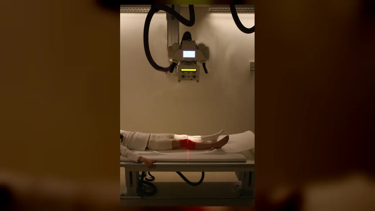
⚡ Everyday Impact SparkReveals broken bones and internal organs clearly without a single surgical cut
🤯 Mind-Blowing Fact • Grade 1
Taking a digital X-ray picture takes less than one second—faster than you can say cheese!
✨ Word Sparks (Grade 1 Glossary)
Tap 🔊 to hear
X-ray
an invisible energy beam that pictures bones
lead apron
a heavy vest that blocks extra rays
cast
a hard wrap that helps broken bones heal
🪝
How can a special camera take a picture of your bones right through your skin?
1
Click Goes the Bone Camera
Imagine falling off your bike and hurting your wrist! Inside a quiet hospital room, a friendly imaging helper smiles and slides a smooth camera arm over your arm.
2
Seeing Inside Without Hurting
Click! Invisible beams pass right through soft skin in a blink. Hard white bones show up bright and clear on a computer screen.
3
Safe Heavy Blankets
You get to wear a cozy for safety. You do not feel a thing, and doctors see right away if your bone needs a !
2. Why do patients often wear a heavy lead apron during an X-ray?
🤯 Mind-Blowing Fact • Grade 2
Wilhelm Roentgen discovered X-rays by surprise in 1895 and took the very first human X-ray photo of his wife's hand!
✨ Word Sparks (Grade 2 Glossary)
Tap 🔊 to hear
detector
a digital plate that catches X-ray pictures
fracture
a crack or break inside a bone
calcium
a natural mineral that makes bones strong
🪝
How do hospital imaging experts look inside lungs, joints, and bones without making a single scratch?
1
Calm Guides in the Imaging Room
When you twist an ankle at recess, nobody can tell just by looking if a bone is cracked. Step into the radiology suite, where a calm specialist welcomes you, explains every step, and gently positions your foot on a flat digital .
2
How Invisible Rays Paint Pictures
From behind a clear safety window, the technologist presses a button to send a tiny burst of X-rays. Soft muscles let most rays pass through, appearing gray, while dense in bones blocks the rays and shines bright white.
3
Donut Scanners and Giant Magnets
Many imaging specialists also run donut-shaped CT scanners and tunnel-like MRI machines that use strong magnets instead of X-rays. Clear images help doctors mend and check lungs right away.
1. Why do bones look bright white while muscles look gray on an X-ray?
2. Which imaging machine uses strong magnets instead of X-rays?
🤯 Mind-Blowing Fact • Grade 3
An MRI magnet is so strong that metal wheelchairs, keys, and coins are strictly banned from entering the scan room!
✨ Word Sparks (Grade 3 Glossary)
Tap 🔊 to hear
anatomy
the structure of bones and organs inside bodies
collimator
shutters that narrow an X-ray beam safely
radiologist
a doctor trained to read medical images
🪝
Why must an imaging specialist know every single bone in the human skeleton like a map?
1
Blending Anatomy With High-Tech Cameras
Picture trying to photograph a tiny hairline crack hidden inside one of the twenty-seven bones in a human hand! In hospital imaging suites, technologists combine deep knowledge of human with gentle patient care. They angle both the camera tube and the patient's joint with millimeter precision so hidden injuries never hide behind overlapping bones.
2
Inside the Electromagnetic Spectrum
Just like visible sunlight and radio waves, X-rays belong to the electromagnetic spectrum, except their wavelengths are much shorter and carry higher energy. When a controlled beam crosses the body, air-filled lungs look dark, soft tissues appear in shades of gray, and mineral-rich bones stand out crisp and white.
3
Portable Carts and 3D Slices
Not every patient can walk to the imaging department. Technologists wheel battery-powered portable X-ray units straight into emergency trauma bays and newborn nurseries. Others operate computed tomography, or CT scanners, which spin an X-ray ring around the body to build detailed 3D cross-sections.
4
Smart Shielding and Low Doses
Patient safety always comes first. Technologists narrow the light-guided beam to the exact injured spot and dial in the lowest radiation dose needed to capture a sharp, diagnostic image for the doctor.
1. How do lungs filled with air look on a chest X-ray?
2. Why do technologists use a collimator before taking an X-ray?
🤯 Mind-Blowing Fact • Grade 4
A modern multi-slice CT scanner can spin around a patient three times per second and image the entire chest in less than five seconds!
✨ Word Sparks (Grade 4 Glossary)
Tap 🔊 to hear
modality
a specific type of medical imaging method
fluoroscopy
real-time moving X-ray video of internal organs
photon
a tiny packet of light or electromagnetic energy
🪝
How do modern hospital imaging teams turn invisible waves and magnetic fields into crystal-clear 3D maps of the body?
1
First Eyes in Trauma and Orthopedics
Sirens fade outside the emergency entrance as paramedics wheel in an injured soccer player. Within minutes, a medical imaging technologist glides a ceiling-mounted X-ray tube into place, comforts the nervous athlete, and captures razor-sharp views from two perpendicular angles. Because flat two-dimensional images can flatten depth, taking both front-to-back and side-view projections reveals exact bone alignment for orthopedic doctors.
2
Digital Detectors and the ALARA Principle
Gone are the darkrooms and chemical film tanks of the past; modern flat-panel digital detectors convert incoming X-ray into electronic signals that appear on high-resolution monitors in seconds. Every technologist practices a golden safety rule called ALARA—As Low As Reasonably Achievable. By adjusting kilovoltage for beam penetration and milliamperes for beam quantity based on a child's or adult's size, they minimize exposure while preserving crisp detail.
3
Beyond X-Rays: CT, Fluoroscopy, and MRI
Experienced technologists often specialize in advanced imaging modalities. In computed tomography (CT), a rotating gantry captures hundreds of thin cross-sectional slices in a single breath-hold to spot internal bleeding or appendicitis. In , live moving X-ray video helps surgeons guide tiny catheters through blood vessels. Meanwhile, magnetic resonance imaging (MRI) aligns hydrogen atoms inside body water using superconducting magnets and radiofrequency pulses to map brain tissue and knee ligaments without any ionizing radiation.
4
Teamwork Between Technologist and Physician
Capturing a clear scan requires calm communication, physics mastery, and creative problem-solving—especially when helping hurt patients hold perfectly still without worsening pain. Once images pass quality review, technologists transmit them over secure hospital networks to board-certified radiologists, turning invisible physics into fast, non-invasive healing.
1. Why do technologists usually take at least two X-ray views from different angles?
2. What does the safety rule ALARA remind technologists to do?
🤯 Mind-Blowing Fact • Grade 5
Superconducting MRI magnets are cooled with liquid helium to minus 269 degrees Celsius—just four degrees warmer than absolute zero in deep space!
✨ Word Sparks (Grade 5 Glossary)
Tap 🔊 to hear
attenuation
gradual weakening of energy passing through matter
voxel
a tiny 3D cube in digital scans
dosimeter
a badge measuring personal radiation exposure levels
🪝
How does applied quantum physics allow medical imaging specialists to peer through solid bone, beating hearts, and delicate neural pathways?
1
Where Applied Physics Meets Compassionate Care
Inside a modern diagnostic imaging center, high-voltage generators accelerate electrons across a vacuum tube at nearly half the speed of light, slamming them into a rotating tungsten anode to produce tightly focused X-ray photons. Operating this sophisticated equipment requires a certified specialist fluent in both radiation physics and three-dimensional human anatomy. Equally vital is calm bedside empathy: guiding anxious children, elderly stroke patients, or immobilized trauma victims through exact positioning maneuvers without causing discomfort.
2
Differential Attenuation and Digital Radiography
Every radiograph relies on differential —the physical principle that tissues of varying atomic number and density absorb X-ray photons at different rates. Calcium-rich cortical bone (atomic number 20) strongly absorbs photons via the photoelectric effect, leaving unexposed white shadows on the digital cesium-iodide detector, whereas low-density lung alveoli allow photons to pass unimpeded. Technologists fine-tune tube potential (kilovolt peak, kVp) to control image contrast and tube current-time product (milliampere-seconds, mAs) to eliminate quantum mottle.
3
Helical Computed Tomography and Interventional Suites
When emergency physicians suspect a pulmonary embolism, cranial hemorrhage, or complex pelvic fracture, CT technologists operate helical scanners whose X-ray tubes and multi-row detector arcs whirl continuously around a sliding table. Mathematical reconstruction algorithms assemble thousands of measurements into sub-millimeter 3D . In interventional angiography suites, sterile-gowned technologists run C-arm fluoroscopes while injecting iodinated contrast media, illuminating coronary arteries in real time as cardiologists open blocked vessels with tiny stents.
4
Nuclear Spin and Superconducting MRI
Cross-training into magnetic resonance imaging introduces an entirely non-ionizing realm of physics. Cooled by liquid helium to minus 269 degrees Celsius, a 1.5- or 3-Tesla superconducting coil aligns spinning hydrogen protons throughout the patient's body water. Timed radiofrequency pulses tip those protons out of alignment; as they relax back, receiver coils record faint radio echoes that computers Fourier-transform into exquisite maps of spinal discs, cartilage, and brain white matter.
5
Dosimetry, Ethics, and Diagnostic Stewardship
Registered through the American Registry of Radiologic Technologists (ARRT), professionals wear personal badges to monitor occupational exposure while enforcing time, distance, and lead shielding protocols. By tailoring pediatric exposure charts, screening every MRI visitor for ferromagnetic implants, and verifying anatomical side markers on PACS networks, technologists safeguard patients while illuminating hidden pathways to recovery.