🧬 HUMAN BODY
🧬 Nervus Opticus (Cranial Nerve II) • Visual Pathway Tract
VA Science & Health SOL 1.1, 3.1, 4.2, 5.1 (Cells, Organs & Body Systems)
Optic Nerve
High-Speed Video Cable to Your Brain
Grade 5 • Advanced • 310 words
🩺 Body System & LocationCentral Nervous System • Back of each eyeball through skull socket to visual brain
🔬 Size / Cell CountAbout 2 in (5 cm) long & 0.16 in (4 mm) thick • Bundles ~1.2 million nerve fibers per eye
⚙️ Core JobStreams every color, shape, and motion signal from your retina to your brain
⚡ Body SuperpowerWhere it exits the eye sits a natural 'blind spot' that your clever brain paints over invisibly!
🤯 Mind-Blowing Fact • Grade 1
Even though your eyes sit in the front of your head, your optic nerves carry picture news all the way to the VERY BACK of your brain!
✨ Word Sparks (Grade 1 Glossary)
Tap 🔊 to hear
optic
Having to do with eyes and seeing
cord
A neat bundle of thin fibers together
stream
To flow smoothly without stopping
🪝
How does a picture inside your eye reach the back of your brain?
1
Plugged Into Your Brain!
Your eyes catch bright pictures all day long! Yet your eyes cannot understand what they see all alone. Behind each eye plugs in a fast called the nerve!
2
Streaming Live Movies
Over a million tiny nerve threads pack inside that smooth . They zip color and shape sparks straight back into your brain!
3
See It in a Flash!
Once your brain gets the news, you shout, 'Look, a butterfly!' All of that happens faster than one blink!
1. What job does the optic nerve do behind each eyeball?
2. About how many tiny nerve threads pack inside just one optic nerve?
🤯 Mind-Blowing Fact • Grade 2
French scientist Edmé Mariotte shocked King Charles II's court in 1668 by using the optic nerve's blind spot to make a person's head 'disappear' when viewed with one closed eye!
✨ Word Sparks (Grade 2 Glossary)
Tap 🔊 to hear
exit
A doorway where things travel outward
overlap
Covering part of the exact same space
automatically
Working smoothly without needing your help
🪝
Did you know each of your eyes has a tiny 'blind spot' hiding right in plain view?
1
Like a TV Video Cable
Think of how a video game console needs a cable to show bright games on a television screen! Behind each eyeball, a shoelace-thick cable called the optic nerve carries over a million sight wires straight into your brain.
2
The Secret Blind Spot
Right where that thick cable the back wall of your eye, there is zero room for light-catching rod or cone cells! That tiny exit circle creates a natural blind spot in every human eye.
3
Your Brain Fills It In
Why don't you notice two empty holes floating in front of you? Your two eyes their views, and your smart brain paints in matching colors !
1. Why does every human eye have a tiny natural blind spot?
2. Why don't you see blank holes in the world from your blind spots?
🤯 Mind-Blowing Fact • Grade 3
Animals with eyes on the sides of their heads like horses and rabbits cross almost ALL fibers at their optic chiasm, while front-eyed humans cross almost exactly fifty percent for 3D depth!
✨ Word Sparks (Grade 3 Glossary)
Tap 🔊 to hear
chiasm
X-shaped crossroads where nerve fibers trade sides
occipital
Back brain lobe processing everything you see
depth
Judging how near or far objects stand
🪝
Why do the optic nerves from your left and right eyes meet at an X-shaped crossroads under your brain?
1
Cranial Nerve Number Two
Doctors number the twelve head nerves from front to back: number one smells cookies, and number two—your optic nerve—delivers the entire visual world! Stretching about five centimeters from the back of each eyeball through a bony tunnel in the skull, each cord bundles roughly 1.2 million myelinated axons.
2
Meeting at the Optic Chiasm
Deep behind your nose and just below your hypothalamus, your left and right optic nerves meet at an X-shaped crossroads called the optic (named after the Greek letter chi, X!). Half the fibers from each eye cross over to the opposite side while the other half stay put.
3
Building 3D Depth Vision
Why trade half their wires? Crossing brings matching views from both eyes together on the same side of your brain—letting your visual cortex judge 3D when you catch a basketball!
4
Destination: Occipital Lobe
After a quick relay stop in the thalamus, signals reach the lobe at the very back of your head, turning sparks into recognizable faces and words!
1. What happens at the X-shaped optic chiasm beneath your brain?
2. Which lobe at the very back of your brain finally turns optic nerve signals into clear sight?
🤯 Mind-Blowing Fact • Grade 4
Your intraorbital optic nerve is built about five millimeters longer than the straight-line distance across your eye socket, resting in a gentle S-curve so darting eye movements never pull it taut!
✨ Word Sparks (Grade 4 Glossary)
Tap 🔊 to hear
hemisphere
Either the left or right brain half
nasal
Closer to the center nose side
temporal
Closer to the outer temple side
🪝
Why does everything on the LEFT side of your playground end up in the RIGHT half of your brain?
1
Clever Left-Right Traffic Sorting
Stare straight ahead at the center of a soccer field! Light from players on the left half of the field enters BOTH of your eyes—hitting the (nose-side) retina of your left eye and the (temple-side) retina of your right eye. At the X-shaped optic chiasm, nasal fibers cross the midline while temporal fibers stay on their own side. Thanks to that brilliant geometric sorting, every clue from the left half of the world lands neatly in your brain's right , while the right half of the world lands in your left hemisphere!
2
Secretly a Central Brain Tract!
Although everyone calls cranial nerve II a 'nerve,' anatomists love pointing out that your optic nerve is technically a central nervous system white-matter tract! Unlike arm nerves wrapped by Schwann cells, optic nerve axons are insulated by brain oligodendrocytes and wear all three protective meningeal layers bathed in cerebrospinal fluid.
3
Slack for Swiveling Eyes
Glance sharply left, right, up, and down! Because the distance from eye socket to eyeball back is only about 25 millimeters while the intraorbital optic nerve measures roughly 30 millimeters in a gentle S-curve, five millimeters of built-in slack let your eyeballs swivel freely without ever tugging the cable!
4
Relay Through the Thalamus
Beyond the chiasm, fibers continue as optic tracts to the lateral geniculate nucleus of the thalamus, which sharpens focus and fans signals back to the visual cortex.
1. Why is the optic nerve inside your eye socket curved in a gentle S-shape with extra slack?
2. How do doctors know the optic nerve is technically a central nervous system tract rather than an ordinary peripheral nerve?
🤯 Mind-Blowing Fact • Grade 5
In 1704—nearly two hundred years before neuroscientists could trace individual nerve fibers—Isaac Newton used pure geometry to correctly predict that nasal optic nerve fibers cross at the chiasm while temporal fibers stay uncrossed!
How does a four-millimeter biological fiber-optic cable stream ten megabits of compressed neural video per second while feeding its own central blood vessels?
1
Bandwidth of the Retinal Ganglion Highway
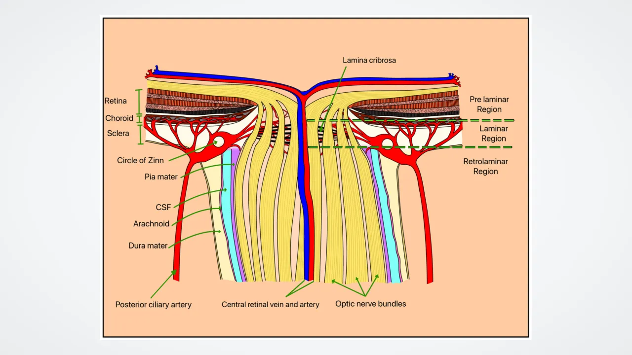
Converging toward the nasal optic disc (papilla), approximately 1.2 million unmyelinated retinal ganglion cell axons thread through a sieve-like collagen mesh in the sclera called the lamina cribrosa. Instantly behind that sieve, oligodendrocytes wrap every fiber in compact myelin—doubling the cable's diameter from 1.5 millimeters at the disc to roughly 4 millimeters inside the orbit and boosting conduction speeds up to fifty meters per second! Biophysical calculations estimate each human optic nerve streams roughly ten megabits per second of edge-and-motion-compressed visual code.
2
Vascular Core and Intracranial Pressure Clues
About twelve millimeters behind the globe, the central retinal artery and vein pierce right into the core of the optic nerve to emerge through the center of the optic disc and nourish the inner retina. Because the subarachnoid space inside the optic nerve's meningeal sheath connects directly with fluid surrounding the brain, neurologists peer through an ophthalmoscope at the optic disc to check whether brain fluid pressure or eye pressure is straining that delicate sieve!
3
Isaac Newton's Brilliant Prediction
Long before modern microscopes could trace axons, physicist Isaac Newton deduced in his 1704 book 'Opticks' that achieving unified single vision from two eyes required partial —crossing of only nasal fibers—at the optic chiasm! Nearly two centuries later, Santiago Ramón y Cajal confirmed Newton's exact geometric prediction under the microscope.
4
Three Destinations Beyond the Chiasm
Passing into the optic tracts, ninety percent of fibers synapse inside the six-layered lateral geniculate nucleus (LGN) of the thalamus before fanning through Meyer's loop and optic radiations to primary visual cortex (V1). Meanwhile, ten percent branch to the midbrain's superior colliculus to steer lightning-fast saccadic eye movements and to the pretectum and suprachiasmatic nucleus for pupil reflexes and daily rhythms!
5
Neuroprotection and Regeneration Frontiers
Because adult central nervous system tracts normally resist regrowing severed axons, vision scientists at leading eye institutes are pioneering gene switches and neurotrophic growth factors aimed at coaxing injured retinal ganglion axons to regenerate all the way back to the thalamus.