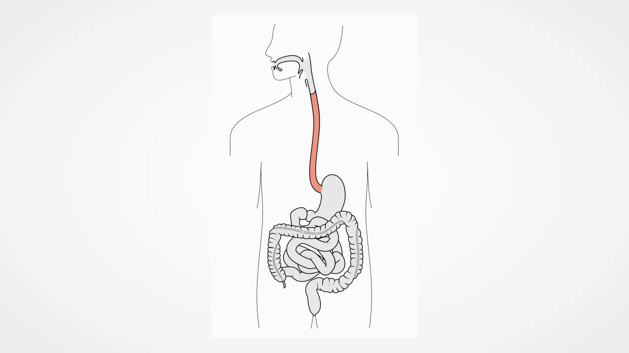
1. How does chewed food move down your esophagus into your stomach?
2. What sits at the bottom of your esophagus to welcome food into the stomach?
🤯 Mind-Blowing Fact • Grade 2
Even if you safely stood on your head (don't try eating upside down!), peristalsis muscle waves are strong enough to push food uphill into your stomach!
✨ Word Sparks (Grade 2 Glossary)
Tap 🔊 to hear
Peristalsis
Traveling wave of muscle squeezes moving food
Valve
One-way doorway keeping liquids moving one direction
Ribbon
Something long, soft, and flat when resting
🪝
How long does it take a bite of pancake to ride the elevator from your throat to your stomach?
1
Hidden Behind Your Windpipe
Swallow a sip of cool water! Right behind your ringed windpipe runs a flat, pink muscular about ten inches long in grown-ups. That food highway is your esophagus. When nothing is passing through, it rests gently flat to save space in your chest.
2
The Wave Called Peristalsis
As soon as a chewed bite enters the top, muscles right behind the bite squeeze tight while muscles ahead of it relax! Scientists call this traveling wave . Liquids zip down in two seconds, while solid bites arrive in about five to eight seconds.
3
Two Smart Gatekeepers
Ring-shaped muscle guard both ends—keeping air out at the top and keeping sour tummy juices down at the bottom!
1. What is the scientific name for the traveling wave of muscle squeezes in your esophagus?
2. Roughly how many seconds does a chewed bite of solid food take to travel down the esophagus?
🤯 Mind-Blowing Fact • Grade 3
Your heart has nothing to do with 'heartburn'—the nickname exists only because your esophagus passes right behind your heart inside your chest!
✨ Word Sparks (Grade 3 Glossary)
Tap 🔊 to hear
Sphincter
Circular muscle ring acting like a drawstring door
Bolus
Smooth, saliva-moistened ball of chewed food
Acid
Sour digestive juice inside your stomach
🪝
Why is the curious burning feeling called 'heartburn' actually happening in your esophagus instead of your heart?
1
Threading Through a Crowded Chest
Your esophagus takes an exciting route from throat to belly! Starting right behind your trachea in the neck, it travels straight down behind your heart and lungs, then slips through a special muscular doorway right in your diaphragm to join the top of your stomach.
2
Two Layers of Muscle Teamwork
How does peristalsis glide so smoothly? Two muscle layers wrap the esophagus in different directions! An inner layer forms tight rings that pinch behind the chewed food ball (), while an outer lengthwise layer shortens ahead of the bite to pull the tube gently over it.
3
Switching From Choice to Automatic
Starting a swallow uses voluntary muscle at the very top, yet halfway down, smooth muscle takes over on 100 percent automatic pilot!
4
The Mystery of 'Heartburn'
At the bottom, the lower esophageal closes tightly so sour stomach stays put. If that ring briefly lets a splash of acid touch the esophagus right behind your heart, people feel 'heartburn'—even though their heart is completely fine!
1. Through which breathing muscle must your esophagus pass to reach your stomach?
2. Why is an acid splash at the bottom of the esophagus nicknamed 'heartburn'?
🤯 Mind-Blowing Fact • Grade 4
If a sticky bite of toast pauses halfway down your chest, stretch sensors in your esophagus launch a silent backup 'secondary wave' automatically without another swallow!
✨ Word Sparks (Grade 4 Glossary)
Tap 🔊 to hear
Striated
Striped fast-acting muscle tissue like arm muscles
Mucosa
Moist, protective inner lining of digestive tubes
Reflux
Backward splash of stomach juice upward
🪝
Why does the top of your esophagus use arm-style muscle while the bottom uses stomach-style muscle?
1
A Three-Zone Muscle Transition
Examine the muscular wall (muscularis externa) of the twenty-five-centimeter human esophagus under a microscope, and you will spot a remarkable relay handoff! The top third is built of fast-contracting skeletal muscle—the same type found in your arms and tongue—so your swallow begins crisp and quick. The middle third blends skeletal and smooth muscle fibers side by side, while the bottom third is 100 percent involuntary smooth muscle, handing control seamlessly over to your digestive tract's own built-in nerve web!
2
Primary and Secondary Sweeps
What happens if a sticky bite of peanut-butter toast lingers halfway down? Stretch sensors built right into the esophageal wall detect the bulge and automatically launch a 'secondary peristaltic wave'—without you even needing to swallow again—sweeping the last crumb cleanly into the stomach!
3
Folded Inner Armor
Unlike the stomach below it, which absorbs water or alcohol and secretes harsh acid, the esophagus is strictly a transport corridor. Its inner folds into lengthwise pleats when empty so it can stretch wide for big bites, and wears dozens of stacked layers of tough squamous cells lubricated by mucus glands to resist friction.
4
Diaphragm Pinch-Helper
Right where the lower esophageal sphincter meets the stomach, muscle loops of your diaphragm wrap around the outside like an extra belt—tightening every time you inhale so chest suction never pulls stomach juices upward!
How can your esophagus coordinate sweeping waves using its own 'second brain' of local neurons?
1
Four Tunics and Absence of Serosa
Like the rest of the gastrointestinal tract, the esophagus is constructed from four concentric tissue tunics: mucosa, submucosa, muscularis propria (inner circular and outer longitudinal layers), and an outer fibrous coat. Unlike abdominal organs suspended in slippery peritoneal serosa, the thoracic esophagus wears a loose connective-tissue that tethers it flexibly inside the mediastinum while still letting it glide smoothly during breathing and neck motion. Its non-keratinized stratified squamous epithelium renews rapidly every week, lubricated by deep submucosal mucous glands.
2
Enteric Nervous System: Your 'Second Brain'
Nestled directly between the inner circular and outer longitudinal muscle coats lies Auerbach's (myenteric) plexus—part of the Enteric Nervous System, a self-contained network of hundreds of millions of gut neurons! When vagal efferents initiate primary peristalsis, excitatory neurons release acetylcholine right behind the bolus to contract circular smooth muscle, while inhibitory neurons ahead of the bolus release (NO) and vasoactive intestinal peptide (VIP) to relax downstream muscle—propelling food at 2 to 4 centimeters per second against pressures up to 100 mmHg.
3
Upper and Lower Sphincter Physiology
Guarding the cervical entrance at vertebra C6, the anatomical Upper Esophageal Sphincter (cricopharyngeus muscle) stays tonically contracted at rest so inspired air enters only the trachea, relaxing for less than a second per swallow. At the gastroesophageal junction (T11), the physiological Lower Esophageal Sphincter pairs intrinsic smooth-muscle tone with external crural diaphragm fibers and the acute Angle of His—forming a flap-like anti-reflux barrier.
4
Kussmaul and Sword-Swallower Endoscopy
How did physicians first peer inside a living human esophagus? In 1868, German internist Adolf Kussmaul enlisted a professional carnival sword-swallower to test a lighted rigid metal tube—pioneering esophagoscopy before modern ultra-thin flexible fiber-optic cameras transformed digestive medicine!
5
High-Resolution Manometry Maps
Today, gastroenterologists map esophageal pressure waves in rainbow-colored contour plots called Clouse plots, diagnosing motility puzzles down to the millimeter.