1. Why does the outside of your cerebrum have so many wrinkles and folds?
2. Which side of your cerebrum sends the signal when you wave your right hand?
🤯 Mind-Blowing Fact • Grade 2
Even though your brain weighs only about two percent of your body, your busy cerebrum uses roughly twenty percent of your body's energy and oxygen!
✨ Word Sparks (Grade 2 Glossary)
Tap 🔊 to hear
hemisphere
one of the cerebrum's two halves
cortex
wrinkled outer thinking layer of brain
neuron
a messenger cell firing tiny sparks
🪝
What happens if you unfold all the bumps and grooves on the top of your brain?
1
A Crumpled Newspaper Trick
Spread a big sheet of newspaper flat on your desk, then crumple it into a ball the size of a grapefruit! Your cerebrum uses that exact folding trick. Accounting for eighty-five percent of your brain's weight, its outer bark—the cerebral —would cover two and a half square feet if ironed flat!
2
Bridging Left and Right
A deep groove divides your cerebrum into left and right . Deep in the middle, a thick cable of nerve fibers called the corpus callosum zips messages back and forth between both halves.
3
Jobs for Every Sense
Whether you solve math puzzles, recognize a friend's voice, taste strawberries, or kick a soccer ball, your cerebrum runs the show!
1. About how much of your total brain's weight belongs to the cerebrum?
2. What connects the left and right hemispheres so both sides of your cerebrum share teamwork?
🤯 Mind-Blowing Fact • Grade 3
Your cerebrum itself has zero pain receptors—meaning it feels tickles or stubbed toes from your whole body, yet feels nothing directly on itself!
✨ Word Sparks (Grade 3 Glossary)
Tap 🔊 to hear
lobe
a major specialized section of cerebrum
gray matter
outer brain layer packed with cell bodies
voluntary
actions you choose to do on purpose
🪝
Why is the seeing center of your cerebrum tucked all the way at the very back of your head instead of behind your eyes?
1
Four Specialized Neighborhoods
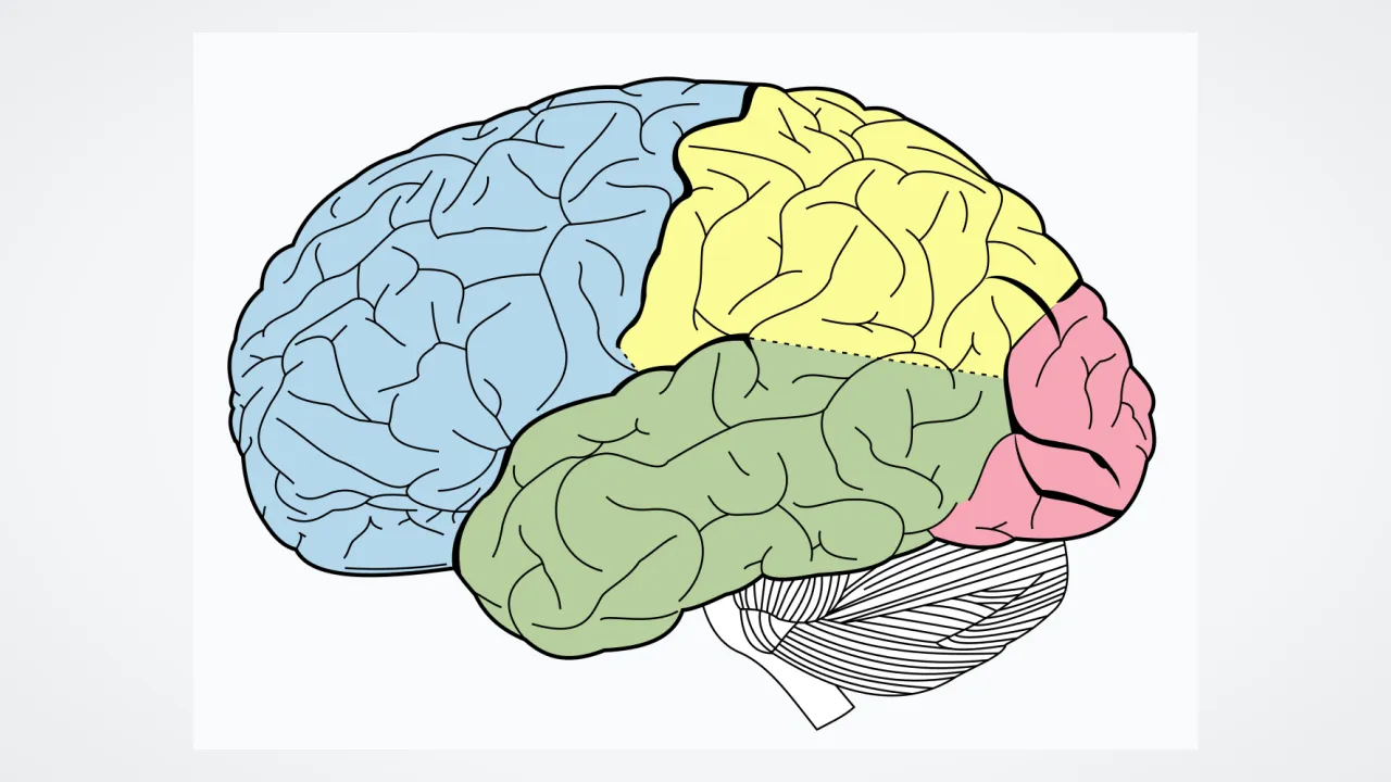
Imagine a bustling city divided into four busy neighborhoods! Each hemisphere of your cerebrum has four major regions called , named after the skull bones protecting them. Even though they look alike on the surface, each lobe specializes in amazing jobs.
2
Frontal and Parietal Lobes
Right behind your forehead sit the frontal —your headquarters for planning, speaking, solving problems, and sending movement commands to your muscles. Behind them sit the parietal lobes, processing touch, warmth, and spatial awareness.
3
Temporal and Occipital Lobes
Above your ears work the temporal , decoding music, spoken words, and memories. And tucked all the way at the back of your head sit the occipital lobes, turning signals from your eyes into vivid 3D color vision!
4
Gray Matter and White Matter
Billions of neuron cell bodies form the thin outer '' cortex, while insulated 'white matter' axons wire distant together at lightning speed.
1. Which lobe at the very back of your cerebrum turns eye signals into sight?
2. Which lobe right behind your forehead handles planning, speaking, and voluntary muscle movement?
🤯 Mind-Blowing Fact • Grade 4
Signals racing along myelinated white-matter highways inside your cerebrum can travel over 250 miles per hour—faster than a Formula 1 race car!
✨ Word Sparks (Grade 4 Glossary)
Tap 🔊 to hear
gyri and sulci
folded ridges and grooves of cortex
synapse
tiny gap where neurons pass messages
neuroplasticity
brain's ability to rewire through learning
🪝
Why do your fingers and lips get way more space on your cerebrum's touch map than your entire back?
1
Ridges, Grooves, and Six Cortical Layers
Peer closely at the cerebrum—known anatomically as the telencephalon—and every hill and valley has a scientific name: raised ridges are called gyri, and folded valleys are called sulci. Only two to four millimeters thick, this outer neocortex stacks roughly 16 billion neurons into six microscopic layers wired by over a hundred trillion synaptic connections!
2
The Surprising Sensory Homunculus
Running across the top of your cerebrum like a pair of headphones sit two matching strips: the motor cortex (firing muscle commands) and the somatosensory cortex (receiving touch). Surprisingly, body parts aren't mapped by physical size—they are mapped by sensitivity and precision! Your fingertips, lips, and tongue claim huge territories of neurons, while your broad back gets only a tiny sliver.
3
Myth-Busting Left Brain vs. Right Brain
You may have heard people claim someone is strictly 'left-brained' for math or 'right-brained' for art. Modern functional MRI scans busted that myth! While language centers like Broca's and Wernicke's areas usually sit in the left hemisphere, creative writing, music, and math light up massive networks across both hemispheres simultaneously via the 200-million-axon corpus callosum.
4
Neuroplasticity in Action
Every time you practice Spanish vocabulary or master skateboard tricks, your cerebrum physically strengthens active —a lifelong superpower called .
1. Why do your fingertips take up much more room on your somatosensory cortex map than your whole back?
2. What have modern brain scans proven about the popular 'left-brained vs. right-brained personality' myth?
🤯 Mind-Blowing Fact • Grade 5
Despite outperforming massive supercomputers at recognizing faces and understanding jokes simultaneously, your entire cerebrum runs on roughly 20 watts of power—less electricity than a standard laptop!
brain regions blending senses into complex thought
🪝
How do 16 billion cortical neurons speaking only in tiny electrical pulses and chemical whispers construct every sight, word, and daydream you experience?
1
Evolutionary Expansion of the Telencephalon
Developing from the embryonic forebrain (prosencephalon), the human cerebrum—or telencephalon—crowns the central nervous system across roughly 1,200 cubic centimeters. What distinguishes primate brains, and humans above all, is gyrification: extensive folding of the two-to-four-millimeter-thick neocortex into gyri and deep fissures so that two-thirds of our cortical surface lies tucked inside sulcal walls. Beneath this six-layered gray-matter mantle course white-matter tracts wrapped in lipid-rich sheaths—association fibers linking lobes within one hemisphere, commissural fibers crossing the corpus callosum, and projection fibers descending via the internal capsule toward the thalamus and spinal cord.
2
Columnar Microcircuits and Functional Localization
In the 1860s and 1870s, clinicians Paul Broca and Carl Wernicke demonstrated that focal damage to specific left-hemisphere regions disrupted speech production (frontal Broca's area) versus language comprehension (temporal Wernicke's area) independently. Later, neurosurgeon Wilder Penfield mapped the primary motor and somatosensory strips bordering the central sulcus, revealing an inverted cortical homunculus disproportionately devoted to hands, vocal muscles, and face. Vertically, the neocortex organizes into millions of microscopic cortical columns—functional modules of pyramidal excitatory neurons and GABAergic inhibitory interneurons.
3
Subcortical Partners: Basal Ganglia
The cerebrum isn't solely outer cortex! Buried deep within cerebral white matter sit the subcortical nuclei of the (caudate, putamen, globus pallidus), which filter cortical motor plans so smooth, purposeful actions launch while unwanted twitches stay quiet.
4
Association Networks and Split-Brain Insights
Unlike primary sensory strips, vast multimodal association cortices across frontal, parietal, and temporal lobes integrate sight, sound, memory, and abstract symbols. Roger Sperry's Nobel-Prize-winning studies of patients whose corpus callosum was surgically separated to stop severe seizures revealed lateralized specialization—dominant left-hemisphere syntax paired with right-hemisphere prosody, spatial holistic processing, and facial recognition.
5
Metabolic Demand and Synaptic Pruning
Consuming roughly 20 watts of continuous metabolic power—the brightness of a dim refrigerator bulb—your cerebrum refines trillions of synapses across childhood and sleep, turning daily curiosity into lifelong mastery.