🧬 HUMAN BODY
🧬 Villi Intestinales & Microvilli • Digestive System
VA Science & Health SOL 1.1, 3.1, 4.2, 5.1 (Cells, Organs & Body Systems)
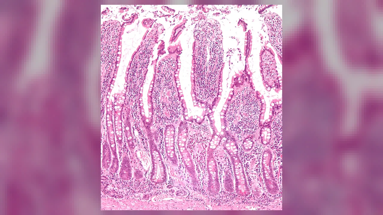
Villi Nutrient Absorbers
Millions of Velvet Finger-Folds Catching Every Single Vitamin
Grade 1 • Early Reader • 76 words
🩺 Body System & LocationDigestive System • Carpeted across the entire inner wall of the small intestine
🔬 Size / Cell Count0.5–1.5 mm tall finger projections • 4–5 million villi multiplying surface area ~600x
⚙️ Core JobCaptures digested sugars, amino acids, vitamins, and fats into blood and lymph vessels
⚡ Body SuperpowerTurns the inside of your skinny gut into a plush velvet carpet as big as a badminton court!
🤯 Mind-Blowing Fact • Grade 1
Millions of microscopic villi make the inside lining of your small intestine look and feel as soft as velvet!
✨ Word Sparks (Grade 1 Glossary)
Tap 🔊 to hear
Villi
Tiny finger-like projections soaking up food nutrients
Velvet
Fabric covered in millions of soft tiny threads
Vitamin
Tiny nutrient helper keeping your body healthy
🪝
Why does a fluffy bath towel dry you so much faster than a flat rain slicker?
1
Like a Fluffy Towel Inside
Touch a soft bath towel! Thousands of tiny cloth loops soak up water fast. Inside your small intestine, millions of tiny finger shapes wave gently just like towel loops! Scientists call them .
2
Catching Every Vitamin
If your gut were smooth inside, lots of good food bits would slide right past! Waving touch every drop of soupy lunch to catch and energy.
3
Straight Into Your Blood
Inside each tiny finger hides a little blood tube ready to carry lunch energy everywhere!
1. How are villi inside your small intestine similar to loops on a fluffy bath towel?
2. What waits right inside every single tiny villus finger?
🤯 Mind-Blowing Fact • Grade 2
A single square inch of your small intestine's inner lining packs roughly 20,000 tiny villi standing shoulder to shoulder!
✨ Word Sparks (Grade 2 Glossary)
Tap 🔊 to hear
Villus
Singular word for just one nutrient-absorbing finger
Microvilli
Ultra-tiny bristles crowning every villus surface cell
Magnify
To zoom in so tiny things look bigger
🪝
What happens when tiny nutrient fingers wear even tinier hairs on top of their heads?
1
Four Million Tiny Fingers
Zoom inside your small intestine with a magnifying camera! Instead of smooth pink walls, you will discover four to five million tiny waving projections called villi, each about one millimeter tall—roughly as thick as a dime's edge.
2
Fuzz on Top of Fingers!
Now zoom in even closer on a single cell covering a ! Sprouting from the top of every cell are thousands of microscopic bristles called . Folds, plus villi, plus microvilli multiply your gut's soaking space hundreds of times!
3
Swaying in the Soup
Tiny muscle strands inside each make them sway gently back and forth, stirring digested food so no nutrient slips past!
1. What crowns the outer cells of each villus to add even MORE nutrient-catching space?
2. Why do tiny muscle strands gently sway your villi back and forth?
🤯 Mind-Blowing Fact • Grade 3
Each tiny villus houses TWO separate highways inside: a blood capillary web for sugars and proteins, plus a central 'lacteal' lymph tube specially built for fats!
✨ Word Sparks (Grade 3 Glossary)
Tap 🔊 to hear
Brush border
Dense carpet of microvilli resembling paintbrush bristles
Lacteal
Central lymph tube inside each villus carrying fats
Stem cell
Builder cell dividing to replace worn-out cells
🪝
Why does every single villus need TWO different kinds of delivery tubes inside its core?
1
Triple Surface-Area Boost
If your small intestine were a plain smooth pipe, its inner surface would barely cover a bathmat—not nearly enough to fuel a growing kid! Three clever layers of folding boost that area enormously: deep circular ridges, millions of finger-like villi, and billions of fuzzy microvilli called the '' because they look like paintbrush bristles under a microscope.
2
Highway One: Blood Capillaries
Covering each villus is a single-cell-thick sheet of absorptive cells. Water-friendly nutrients—such as glucose sugars, protein amino acids, minerals, and vitamins B and C—cross straight through those cells into a fine net of blood capillaries inside the villus!
3
Highway Two: The Lacteal Tube
Digested fats from avocado, butter, or nuts are packaged into droplets too chunky to squeeze through blood capillary walls! Instead, fats enter a special wider green-mapped lymph tube right in the center of each villus, called a .
4
Always Fresh Crew
Busy tucked in shallow valleys between villi grow brand-new absorptive cells every three to five days!
1. Why do digested fats enter the central 'lacteal' tube inside a villus instead of blood capillaries?
2. Why do scientists call the layer of microvilli on villi cells the 'brush border'?
🤯 Mind-Blowing Fact • Grade 4
Discovering that villi cells absorb sodium and glucose side-by-side through a shared turnstile protein led to simple Oral Rehydration Drink packets that have saved over 50 million lives!
✨ Word Sparks (Grade 4 Glossary)
Tap 🔊 to hear
Osmosis
Water naturally following dissolved salts and sugars across cells
Chylomicron
Protein-coated fat parcel entering a villus lacteal
Gluten
Stretchy grain protein found in wheat and rye
🪝
How did one simple discovery about how villi absorb sugar and salt together save tens of millions of children around the world?
1
Multiplying Area Six Hundred Times
Mathematicians marvel at how the small intestine packs vast absorptive territory into a compact belly! Circular plicae folds triple the gut's inner area; finger-like villi multiply that by ten; and roughly 3,000 actin-cored microvilli crowning every single enterocyte multiply it by twenty more—yielding a 600-fold total boost covering over 30 square meters (bigger than a badminton court)!
2
The Two-Rider Turnstile Trick
Villi don't just wait passively for nutrients to drift across. On the brush border sits a remarkable molecular turnstile protein called SGLT1 that refuses to turn unless ONE sodium salt ion and ONE glucose sugar molecule step aboard together! As millions of sodium-glucose pairs ride into enterocytes, water naturally follows right behind them by . Doctors realized that giving sick, dehydrated kids simple clean water mixed with exact pinches of salt and glucose—Oral Rehydration Solution—switches on those villi turnstiles even during tummy bugs, saving tens of millions of young lives globally!
3
Why Lacteals Turn Milky White
Inside enterocytes, fatty acids are reassembled and wrapped in protein coats called before entering the central lacteal—turning the lymph fluid temporarily milky white ('chyle'), which inspired the Latin name lacteal ('milky')!
4
Understanding Celiac Sensitivity
In people with celiac disease, eating wheat protein () confuses immune cells into flattening villi; switching to gluten-free foods lets stem cells regrow tall, healthy villi again!
1. Why does mixing precise amounts of both glucose sugar AND salt into water rehydrate the body faster than plain water during stomach bugs?
2. Where did the central 'lacteal' vessel inside each villus get its 'milky' Latin name?
🤯 Mind-Blowing Fact • Grade 5
Stem cells nestled in Crypts of Lieberkühn rebuild your entire velvet carpet of intestinal villi from bottom to tip every 3 to 5 days—the fastest-renewing tissue lining in your body!
✨ Word Sparks (Grade 5 Glossary)
Tap 🔊 to hear
Cotransport
Moving two different molecules across a cell together
Crypt
Deep stem-cell valley nestled between neighboring villi
Organoid
Tiny 3D lab-grown organ model built from stem cells
🪝
How does an escalator of stem cells deep in intestinal 'crypts' rebuild your entire velvet carpet of villi every four days?
1
Architecture of the Crypt-Villus Axis
Each intestinal villus—tallest and leaf-shaped in the duodenum (~1.5 mm) and tapering into finger-like projections in the ileum (~0.5 mm)—projects from the lamina propria wrapped in a single continuous layer of simple columnar enterocytes sealed together at their apices by tight junctions. That single-cell thickness is deliberate: nutrients cross fewer than thirty micrometers from intestinal lumen to fenestrated blood capillaries or the blind-ended central lymphatic lacteal, while strands of smooth muscle extending from the muscularis mucosae rhythmically milk chyle upward toward the thoracic duct.
2
Active Cotransport and Energetics
Along the basolateral membrane of every enterocyte, Na+/K+-ATPase pumps burn ATP nonstop to keep intracellular sodium low. That steep electrochemical sodium gradient powers secondary active symporters along the apical microvilli—such as SGLT1 for glucose/galactose and PepT1 proton-coupled transporters for di- and tripeptides—while fructose crosses via facilitated GLUT5 channels! Because Robert Crane's 1960 discovery of sodium-glucose remains intact even during cholera toxin exposure, WHO Oral Rehydration Therapy became a cornerstone of global pediatric medicine.
3
Crypts of Lieberkühn: Fastest Cell Renewal
Surrounding the base of every villus plunge tubular valleys called intestinal glands, or of Lieberkühn. At the bottom of each crypt, Lgr5-positive multipotent stem cells—shielded by antimicrobial lysozyme-secreting Paneth cells—divide continuously! Daughter cells ride an upward cellular escalator along the villus flank, differentiating into absorptive enterocytes, mucus-making goblet cells, and hormone-secreting enteroendocrine cells before shedding harmlessly from the villus tip after just three to five days—the fastest epithelial turnover rate in the human body!
4
Gaspare Aselli and Milky Vessels (1622)
Lymphatic absorption remained unknown until 1622, when Italian anatomist Gaspare Aselli observed snow-white 'vene albe et lacteae' (milky veins) threading from post-meal mesentery—discovering lacteals six years before Harvey published blood circulation!
5
Gut Organoids on Microchips
In 2009, biologist Hans Clevers proved a single Lgr5+ stem cell grown in 3D gel self-organizes into miniature villi-and-crypt 'mini-guts'—now placed on microfluidic chips to test safe medicines without animal testing.