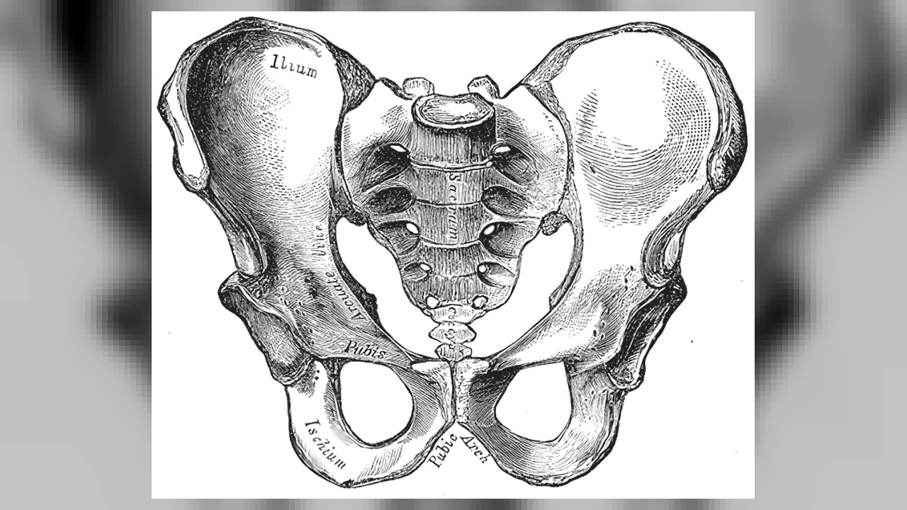
⚡ Body SuperpowerThree separate childhood bones on each side fuse into one indestructible hip socket
🤯 Mind-Blowing Fact • Grade 1
The word 'pelvis' comes straight from the Latin word for a washbowl because its curved bones cradle your lower belly organs like a sturdy bowl!
✨ Word Sparks (Grade 1 Glossary)
Tap 🔊 to hear
Pelvis
Bowl-shaped ring of hip bones at waist
Basin
Wide, rounded bowl with curved sides
Hips
Side joints where legs meet your pelvis
🪝
Put your hands on your hips—what sturdy bone bowl are you touching right now?
1
A Strong Bone Bowl
Stand like a superhero with both hands on your ! Right under your fingers curves a wide ring of bone called your . In Latin, pelvis means '' or 'bowl'!
2
Bridge to Your Legs
Your spine plugs into the back of this sturdy bowl. On the left and right sides, deep round cups hold the tops of your two leg bones.
3
Sitting Bones Underneath
Even sitting in a chair uses your ! Two smooth curved bones at the very bottom rock like chair rails when you sit.
1. What does the Latin word 'pelvis' mean in English?
2. Which two big parts of your skeleton does your pelvis connect together?
🤯 Mind-Blowing Fact • Grade 2
More than thirty different muscles attach to your pelvis, powering everything from standing tall to kicking a soccer ball!
✨ Word Sparks (Grade 2 Glossary)
Tap 🔊 to hear
Fuse
Grow firmly together into one solid bone
Keystone
Center wedge locking a curved arch tight
Cradle
Hold and protect gently from underneath
🪝
Did you know each side of your hip starts out as three separate bones when you are a kid?
1
Three Bones Become One!
Imagine three puzzle pieces slowly growing together until not even a seam shows! All through childhood, your left hip bone and right hip bone are actually made of three separate bones joined by tough cartilage. By your late teens, they into two super-strong hip bones.
2
Completing the Ring
In the front, a springy cartilage pad links the two hip bones together. In the back, the triangle-shaped sacrum at the bottom of your spine wedges tightly between them like the of a stone bridge!
3
Cradling Lower Organs
Inside this protective ring rest your urinary bladder and coils of your intestines, safe from everyday bumps.
1. How many separate childhood bones fuse together to build JUST ONE side of an adult hip bone?
2. Which triangle-shaped bone at the base of your spine locks the back of the pelvic ring together?
🤯 Mind-Blowing Fact • Grade 3
The deep hip socket is called the acetabulum—Latin for 'little vinegar cup'—because ancient Romans thought it looked just like the small bowls they dipped bread into at dinner!
✨ Word Sparks (Grade 3 Glossary)
Tap 🔊 to hear
Ilium
Wide upper wing bone of your hip
Ischium
Curved lower hip bone you sit on
Acetabulum
Deep hip socket holding the femur's ball
🪝
Where do all three parts of your hip bone meet—and why did ancient doctors name that spot a 'vinegar cup'?
1
Ilium, Ischium, and Pubis
Meet the trio of bones that team up on each side of your pelvis! Flaring out at the top like butterfly wings is the —the ridge you feel when you put your hands on your hips. Curving underneath is the L-shaped , your sturdy 'sitting bone.' Bridging across the front is the pubis, where left and right sides meet at a tough fibrocartilage pad called the pubic symphysis.
2
The 'Vinegar Cup' Socket
Right where the , , and pubis fuse together on the outer side sits a deep, smooth cup called the . Ancient Roman anatomists named it 'acetabulum'—Latin for a small vinegar dipping bowl—because its deep socket hugs the round head of the femur so securely!
3
Splitting Weight in Two
Every time you stand or run, weight travels down your single spine into the sacrum, splits left and right across the pelvic arch, and flows down both legs.
4
Blood Cell Powerhouse
Even in grown-ups, the wide flat wings of the hold rich stores of red bone marrow, quietly making fresh blood cells day and night!
1. Which part of the pelvis are you resting your weight on when you sit upright on a hard wooden bench?
2. Where do the ilium, ischium, and pubis all fuse together on the side of each hip?
🤯 Mind-Blowing Fact • Grade 4
Below each hip socket sits the largest hole in any single bone of the human body—the obturator foramen—which shaves off heavy bone weight while giving muscles extra surface to grip!
✨ Word Sparks (Grade 4 Glossary)
Tap 🔊 to hear
Sacroiliac
Super-strong joint linking spine's sacrum to ilium
Symphysis
Tough cartilage pad joining front pubic bones
Gluteus
Powerful hip muscles stabilizing upright walking strides
🪝
Why is a human pelvis shaped like a short, wide bowl while a chimpanzee's pelvis looks like two long, flat paddles?
1
Engineered for Walking on Two Legs
Compare a human skeleton side by side with a chimpanzee skeleton, and the pelvis reveals the secret of upright walking! Chimpanzees have tall, narrow iliac blades lying flat against their backs. If a chimp lifts one foot to step forward, its hips tip sideways unless it sways its whole upper body! In humans, shorter, broader iliac wings wrap forward around the sides, repositioning the medius and minimus muscles so they hold your pelvis level every time one foot leaves the ground.
2
A Self-Locking Roman Arch
At the back of the pelvis, the wedge-shaped sacrum joins the two iliac bones at the (SI) joints, strapped tight by the strongest ligaments in the human body! Just like the central keystone of a Roman stone arch, downward body weight actually wedges the sacrum tighter into the ring.
3
The Obturator Window
Look below the acetabulum on each side and spot a large oval opening—the obturator foramen! Covered mostly by a tough membrane where hip-rotator muscles attach, this window trims excess bone weight without sacrificing ring strength.
4
True Pelvis and Muscular Floor
Stretched across the bottom outlet of the pelvic basin is a supportive muscular hammock—the pelvic floor—cradling urinary and digestive organs against gravity.
1. How do wrap-around human iliac wings help you walk smoothly without swaying side to side?
2. Why does each hip bone have a large oval opening called the obturator foramen?
🤯 Mind-Blowing Fact • Grade 5
Wrapped across the front of your hip socket is the Y-shaped iliofemoral ligament—the single strongest ligament in the human body, able to hold more than 770 pounds without snapping!
✨ Word Sparks (Grade 5 Glossary)
Tap 🔊 to hear
Innominate
Anatomical name for the fused three-part hipbone
Triradiate
Y-shaped childhood growth cartilage inside hip socket
Labrum
Tough cartilage rim deepening a ball-and-socket joint
🪝
How do Y-shaped growth cartilages, Roman-arch biomechanics, and thirty-six muscle attachments make the pelvis the structural crossroads of the human body?
1
Osteology of the Pelvic Girdle
Forming the rigid bony ring of the lower trunk, the adult bony pelvis (pelvis ossea) unites four bones: the paired (coxal) bones anterolaterally, the five-segment fused sacrum posteriorly, and the coccyx inferiorly. Throughout childhood, each coxal bone develops from three primary endochondral ossification centers—the superior ilium, posteroinferior ischium, and anteromedial pubis—meeting at a Y-shaped cartilage inside the acetabular cup that ossifies completely between ages 15 and 17.
2
Closed-Ring Load Transmission and Ligaments
Biomechanically, no isolated beam in the pelvis acts alone; instead, closed-ring geometry distributes loads across two intersecting arches. Axial force descending through L5 enters the sacral promontory, traverses the auricular sacroiliac joints—stabilized by massive interosseous and posterior sacroiliac ligaments—and bifurcates along the arcuate lines of the ilia to either the acetabula (standing) or the ischial tuberosities (sitting), while the fibrocartilaginous pubic symphysis acts as an anterior tension tiebeam resisting ring separation!
3
Ball-and-Socket Stability at the Acetabulum
Deepened around its rim by a fibrocartilaginous collar—the acetabular —and reinforced externally by the spiraled iliofemoral ligament of Bigelow (capable of withstanding over 350 kilograms of tension!), the hip joint sacrifices a fraction of shoulder-like mobility in exchange for rock-solid weight-bearing stability.
4
Anthropological Clues and Marrow Reserves
Anatomists divide the basin along the pelvic brim into the superior false (greater) pelvis supporting abdominal viscera and the inferior true (lesser) pelvis housing pelvic organs above the levator ani muscular diaphragm. Because human bipedalism narrowed the iliac distance while large-brained human infants required an adequate birth canal, adult female pelves typically exhibit a wider subpubic angle (>80 degrees) and rounder pelvic inlet than male pelves.
5
Clinical Significance in Medicine
Rich in lifelong red marrow within the posterior iliac crest, the pelvis also provides hematologists their safest, most accessible site for diagnostic bone-marrow sampling.