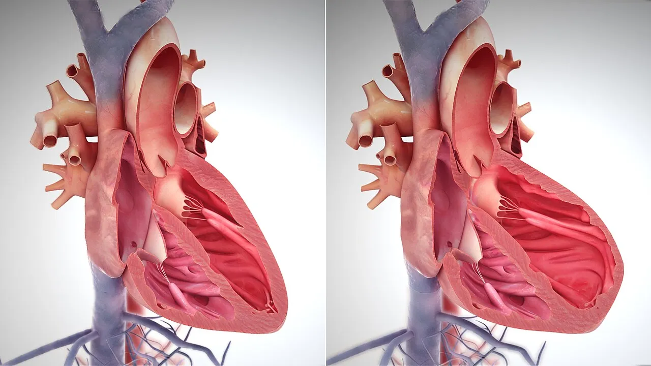
1. Where are the left and right ventricles located inside your heart?
2. Where does the right ventricle send blood?
🤯 Mind-Blowing Fact • Grade 2
Every single day, each of your ventricles pumps nearly two thousand gallons of blood—enough to fill a small swimming pool!
✨ Word Sparks (Grade 2 Glossary)
Tap 🔊 to hear
septum
sturdy muscle wall dividing left and right
oxygen-rich
packed full of fresh oxygen from lungs
muscle
body tissue that squeezes to create motion
🪝
Why is the left side of your heart much thicker and stronger than the right side?
1
Side-by-Side Team Players
Place your hand over your chest after jumping jacks—thump, thump, thump! Each strong beat comes mainly from your left and right ventricles squeezing together at the bottom of your heart. A sturdy wall keeps their two blood streams separate.
2
A Short Trip Next Door
Your right ventricle has an easy neighbor job. It gently pushes used, oxygen-poor blood just a few inches into your spongy lungs to grab fresh oxygen.
3
A Long Highway Journey
Your left ventricle has a giant job—blasting blood up to your brain and down to your feet! That is why its wall grows three times thicker.
1. Why does the left ventricle have a muscle wall three times thicker than the right ventricle?
2. What keeps blood in the left ventricle from mixing with blood in the right ventricle?
🤯 Mind-Blowing Fact • Grade 3
Cardiac muscle inside your ventricles is completely tireless—unlike leg muscles that get sore after sprinting, ventricular muscle beats billions of times without ever taking a vacation!
✨ Word Sparks (Grade 3 Glossary)
Tap 🔊 to hear
systole
pumping phase when heart ventricles squeeze blood out
diastole
relaxing phase when ventricles fill with blood
pulmonary
science word meaning connected to the lungs
🪝
How can two chambers beat at the exact same moment while pumping to totally different places?
1
Twin Pumps, One Rhythm
Press two fingers gently to your wrist and feel a steady pulse! Every pulse wave begins down in your heart's lower chambers, the right and left ventricles. Separated by the thick interventricular septum, both chambers fill and squeeze in synchronized harmony.
2
Gentle Pressure for Delicate Lungs
Why doesn't the right ventricle squeeze with maximum force? Lung air sacs are wrapped in paper-thin capillaries! Your right ventricle uses a thinner muscle wall to send oxygen-poor blood smoothly through the artery without overwhelming delicate lung tissue.
3
High-Pressure Launch From the Left
Once freshly oxygenated blood returns to the left side, the thick, circular left ventricle contracts powerfully, launching blood into the giant aorta fast enough to climb against gravity to your brain.
4
Fill and Squeeze Phases
Doctors call the relaxing fill-up phase and the powerful squeezing launch —repeating over seventy times a minute while you read this sentence!
1. Why is it helpful that the right ventricle pumps at lower pressure than the left ventricle?
2. What do doctors call the phase when your ventricles relax to fill back up with blood?
🤯 Mind-Blowing Fact • Grade 4
Even though the left ventricle builds five times higher pressure than the right ventricle, both chambers MUST pump the exact same volume per minute so blood never backs up in either loop!
✨ Word Sparks (Grade 4 Glossary)
Tap 🔊 to hear
myocardium
thick muscular middle wall of the heart
stroke volume
amount of blood pumped by one heartbeat
Purkinje fibers
fast electrical wires triggering bottom-up heart squeezes
🪝
Why do your heart's ventricles twist like someone wringing water out of a towel with every beat?
1
Spiral Myocardium Architecture
Wring a wet washcloth with both hands—twisting squeezes out far more water than simply clapping your palms flat! Evolution engineered your ventricular (heart muscle) the exact same way. Spiraling layers of cardiac muscle wrap diagonally around both ventricles down to the pointed bottom tip, called the apex, so every contraction twists upward from bottom to top.
2
Pulmonary Loop Versus Systemic Loop
Although the right ventricle looks crescent-shaped in cross-section with walls just three to five millimeters thick, and the circular left ventricle boasts walls ten to fifteen millimeters thick, both eject the exact same —roughly seventy milliliters per beat! The right drives the low-resistance pulmonary circuit at about 25 millimeters of mercury pressure, while the left drives the body-wide systemic circuit near 120 millimeters of mercury.
3
Bottom-Up Electrical Spark
Why squeeze from the bottom tip upward? Exit doors to the pulmonary artery and aorta sit at the top of the ventricles! Insulated electrical highways called the bundle of His and zip signals straight to the bottom apex first, squeezing blood cleanly upward like toothpaste out of a tube.
4
Scaling Up During Exercise
When you race across a basketball court, your ventricles beat faster and contract more forcefully, boosting cardiac output from five liters per minute up to twenty liters or more!
1. Why do Purkinje fibers trigger ventricular contraction starting at the bottom apex first?
2. How does the amount of blood pumped per beat compare between the right and left ventricles?
🤯 Mind-Blowing Fact • Grade 5
More than one-third of every muscle cell inside your heart's ventricles is packed solid with mitochondria—giving cardiac muscle over ten times the power-plant density of ordinary arm muscle!
rule that stretched ventricles automatically squeeze harder
ejection fraction
percentage of chamber blood pumped out per beat
🪝
How do two asymmetrical chambers wired in series pump 7,000 liters daily at a fivefold pressure difference without ever falling out of sync?
1
Two Pumps Wired in Series
Inside the pericardial sac behind your sternum, the human heart operates as two positive-displacement pumps connected in series yet fused into one organ. The thin-walled right ventricle wraps like a crescent pocket around the cylindrical, thick-walled left ventricle. While the right chamber generates peak systolic pressures around 25 mmHg to perfuse nearby alveolar capillaries, the left ventricle reaches roughly 120 mmHg to overcome the vast peripheral resistance of systemic arteries—all while matching an identical resting cardiac output of five liters per minute.
2
Helical Myocardial Band and Torsion
Cardiomyocytes inside ventricular walls link end-to-end via studded with gap junctions that pass electrical ions instantaneously from cell to cell. Arranged in counter-wound subendocardial and subepicardial helices, these muscle sheets twist the cardiac apex counterclockwise during systole to eject roughly sixty percent of end-diastolic blood (), then untwist rapidly during early diastole—creating gentle suction that draws fresh atrial blood inward!
3
His-Purkinje High-Speed Conduction
After pausing briefly at the atrioventricular node so upper chambers finish emptying, impulses race down the interventricular septum along the bundle of His (described by Swiss cardiologist Wilhelm His Jr. in 1893) and fan out through Purkinje fibers (discovered by Jan Evangelista Purkyně in 1839) at four meters per second, firing papillary muscles and apex myocardium in milliseconds.
4
The Frank-Starling Law of Self-Matching
What stops even a one-milliliter mismatch from flooding the lungs? Discovered by Otto Frank and Ernest Starling, the Frank-Starling mechanism ensures that whichever ventricle receives slightly more incoming blood stretches its sarcomeres further and automatically contracts more forcefully on the very next beat!
5
Mitochondrial Endurance Engines
To sustain 2.5 billion lifetime contractions without fatigue, mitochondria occupy nearly thirty-five percent of every ventricular cardiomyocyte's volume—the highest energy-factory density of any muscle in your body.