1. What is the main job of the two atria at the top of your heart?
2. How many atria does one human heart have?
🤯 Mind-Blowing Fact • Grade 2
Each atrium has a wrinkly little flap on the outside called an auricle—named because early doctors thought it looked just like a puppy's floppy ear!
✨ Word Sparks (Grade 2 Glossary)
Tap 🔊 to hear
auricle
wrinkly ear-shaped pouch on each atrium
vein
blood vessel carrying blood back into atria
rhythm
a regular, steady repeating beat
🪝
Why are your heart's top rooms named after the sunny front courtyard of an ancient Roman house?
1
Entrance Halls of the Heart
Step through the front doorway of a busy train station! Long ago, Romans called a home's front entrance hall an atrium. Doctors borrowed that exact name for your heart's two upper rooms—together called the atria—because returning blood enters there first!
2
Two Sides, Two Visitors
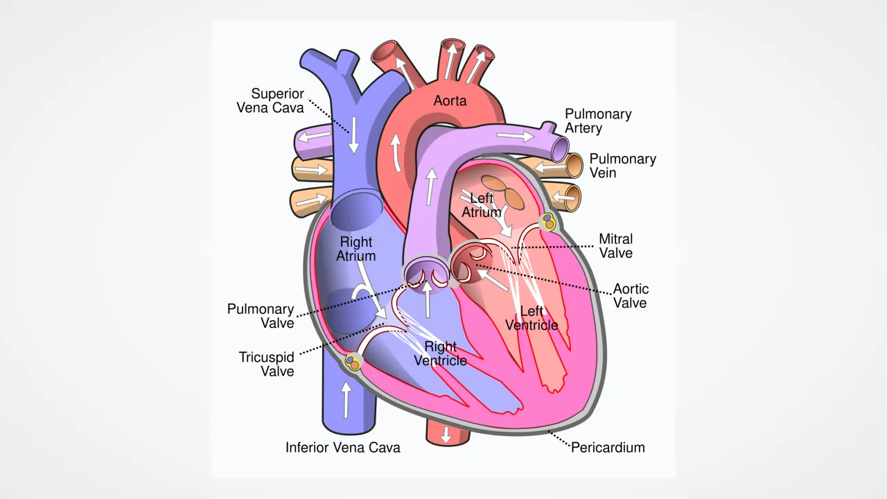
Your right atrium welcomes tired, oxygen-poor blood returning from your arms, legs, and head. Meanwhile, your left atrium welcomes bright, oxygen-rich blood arriving fresh from your lungs.
3
Thin Walls, Quick Squeeze
Because the atria only push blood right downstairs into the ventricles below, their walls stay thin and stretchy. Plus, your right atrium holds the natural pacemaker that starts every single heartbeat!
1. Where does blood entering the LEFT atrium come from?
2. Why do the atria have much thinner muscle walls than the ventricles?
🤯 Mind-Blowing Fact • Grade 3
Before you were born, a tiny doorway called the foramen ovale stayed open between your right and left atria because your mom's placenta supplied oxygen before your lungs took their first breath!
✨ Word Sparks (Grade 3 Glossary)
Tap 🔊 to hear
vena cava
giant vein bringing body blood into right atrium
sinoatrial node
natural pacemaker cell cluster inside right atrium
atrial kick
final gentle squeeze topping off the ventricles
🪝
How do your heart's upper chambers keep blood flowing smoothly instead of stopping and starting?
1
Stretchy Waiting Lounges
Imagine cars trying to merge onto a highway if the on-ramp slammed shut every second! While your lower ventricles squeeze shut to pump blood out, veins keep pouring returning blood nonstop into your stretchy upper chambers, the right and left atria.
2
Seven Big Entry Doors
Three major vessels empty dark, oxygen-poor blood into the right atrium: the superior from your upper body, the inferior vena cava from your lower body, and the coronary sinus from the heart muscle itself. Across the wall, four pulmonary veins pour bright oxygen-rich blood into the left atrium.
3
The Helpful Atrial Kick
As soon as the ventricles relax, doors swing open and about eighty percent of atrial blood glides straight downstairs by itself. Then both atria contract in a quick '' to pack in the final twenty percent!
4
Home of the Sinoatrial Node
High in the right atrium wall sits a tiny bundle of special cells called the —your heart's natural pacemaker that sparks sixty to one hundred beats every minute.
1. Why are stretchy atria important while the lower ventricles are busy pumping?
2. Where is your heart's natural pacemaker—the sinoatrial (SA) node—located?
🤯 Mind-Blowing Fact • Grade 4
Your sinoatrial node inside the right atrium is so self-reliant that even if every nerve from the brain were disconnected, the heart would keep beating steadily on its own!
chemical messenger carried through blood to organs
🪝
Did you know your heart's atria also make a hormone that talks directly to your kidneys?
1
Reservoir, Conduit, and Booster Pump
Sprint across a track, and your heart rate doubles in seconds! Cardiologists describe the paired atria—walled off from each other by the —as three-in-one marvels. With walls just two millimeters thick, they serve as elastic reservoirs while ventricles contract, open conduits when valves swing wide, and booster pumps whose 'atrial kick' supplies up to thirty percent of ventricular filling during intense exercise.
2
Comb-Like Pectinate Muscles
Peek inside the right atrium and its ear-shaped appendage, and you will see parallel muscular ridges that look just like the teeth of a comb! Called , these ridges strengthen the thin atrial wall so it can stretch wide and contract crisply without ballooning out of shape.
3
Setting the Tempo From the SA Node
Near the entrance of the superior vena cava sits a banana-shaped strip of specialized cardiomyocytes called the sinoatrial (SA) node. Unlike ordinary muscles that wait for brain commands, SA node cells spontaneously fire electrical waves across both atria first, making them squeeze fractionally ahead of the ventricles.
4
Secret Hormone Messaging
Your atria are even part of the endocrine system! When extra blood volume stretches atrial walls, atrial muscle cells release a called atrial natriuretic peptide (ANP), signaling your kidneys to let go of extra salt and water so blood pressure stays balanced.
1. When does the 'atrial kick' become extra important for your body?
2. What happens when extra blood stretches the walls of your atria?
🤯 Mind-Blowing Fact • Grade 5
Seconds after a baby takes its very first breath at birth, shifting blood pressures automatically close a secret trapdoor between the right and left atria—rerouting blood through the lungs for the rest of life!
✨ Word Sparks (Grade 5 Glossary)
Tap 🔊 to hear
Bachmann's bundle
fast muscle pathway syncing right and left atria
fossa ovalis
sealed oval dimple marking fetal atrial doorway
preload
volume of blood stretching ventricles before pumping
🪝
How did two landmark 20th-century discoveries reveal that the heart's thinnest chambers serve as both electrical conductor and endocrine gland?
1
Hemodynamic Priming of the Ventricles
Positioned superiorly and posteriorly at the base of the heart, the right and left atria operate at gentle pressures below 12 mmHg, framed by myocardium scarcely two millimeters thick. Despite their modest mass, they convert continuous venous inflow—from the venae cavae and coronary sinus on the right, and four valveless pulmonary veins on the left—into pulsatile ventricular . Across each cardiac cycle, roughly seventy-five percent of blood enters relaxed ventricles passively before coordinated atrial systole delivers the final hemodynamic priming boost.
2
Pacemaker Channels and Bachmann's Bundle
In 1907, anatomists Arthur Keith and Martin Flack identified the microscopic pacemaker tucked near the sulcus terminalis of the right atrium: the sinoatrial node. Powered by 'funny' hyperpolarization-activated ion channels (HCN channels), SA nodal cells drift spontaneously to threshold, launching depolarization waves across the right atrium and racing across a specialized interatrial highway——so both atria contract within milliseconds of each other before pausing 0.1 seconds at the atrioventricular node.
3
Embryonic Remodeling at First Breath
In fetal life, oxygenated placental blood bypasses uninflated lungs by shunting straight from right atrium to left atrium through the flap-guarded foramen ovale. Seconds after a newborn's first breath expands pulmonary vessels, rising left atrial pressure slams the septum primum shut, sealing over into a lifelong thumbprint depression called the .
4
Adolfo de Bold and the Endocrine Heart
For centuries, textbooks viewed the heart strictly as a mechanical pump. In 1981, Argentine-Canadian researcher Adolfo de Bold stunned physiologists by proving that stretched atrial cardiomyocytes secrete granules of atrial natriuretic peptide (ANP)—vasodilating arteries and prompting renal sodium excretion to lower blood pressure.
5
Guarding Rhythm Stability
Understanding atrial conduction around pulmonary vein sleeves also enabled modern cardiologists to restore steady sinus rhythm when disorganized electrical ripples cause atrial fibrillation.