2. What color is the oxygen-poor blood flowing inside your wrist veins?
🤯 Mind-Blowing Fact • Grade 2
At any single moment while you sit reading, nearly two-thirds of all the blood in your entire body is cruising quietly inside your stretchy veins!
✨ Word Sparks (Grade 2 Glossary)
Tap 🔊 to hear
maroon
a deep dark brownish-red color
calf muscle
back lower-leg muscle that squeezes leg veins
reservoir
a roomy storage space holding extra liquid
🪝
If human blood is always red, why do the veins on your wrist look blue?
1
A Clever Trick of Light
Turn your palm upward and spot the veins near your wrist! Blood returning from your hand has already dropped off much of its oxygen, turning deep red. Skin filters incoming sunlight so blue light bounces back to your eyes first—making dark red veins look blue!
2
Wide, Gentle Streams
By the time blood enters your veins, the strong push from your heartbeat has calmed way down. Because pressure is gentle, veins have thinner walls and wider tunnels than arteries.
3
Muscle Pumps and One-Way Doors
Walking flexes your , squeezing deep leg veins like toothpaste tubes while tiny one-way valves catch the blood on every step upward!
1. Why do veins on a fair wrist look bluish to our eyes?
2. How do your leg muscles help your veins when you walk or run?
🤯 Mind-Blowing Fact • Grade 3
Giraffes have extra-tight elastic skin on their long legs—like natural compression socks—plus dozens of heavy-duty vein valves to help blood climb back up to their hearts!
✨ Word Sparks (Grade 3 Glossary)
Tap 🔊 to hear
venule
smallest vein collecting blood straight from capillaries
compression
gentle inward squeezing support around a limb
pulmonary vein
vein bringing fresh oxygen-rich blood from lungs
🪝
Why do soldiers or choir singers sometimes feel dizzy if they lock their knees and stand totally still?
1
Merging Like Tributaries Into a River
Picture tiny mountain brooks joining into streams that pour into one mighty river! After blood trades oxygen inside microscopic capillaries, it gathers into tiny that merge into medium veins and finally empty into two giant trunks—the superior and inferior vena cava.
2
Why Standing Frozen Slows Return
Because venous blood pressure is super low, gravity tugs blood downward into stretchy leg veins when you stand upright. If someone locks their knees without wiggling a muscle, less blood climbs back to the heart for the brain—which is why marching bands bend their knees slightly and wiggle their toes!
3
Breathing Helps Pull Blood Home Too
Even breathing pitches in! Every time you inhale, gentle chest suction helps draw venous blood upward into your right atrium.
4
Pulmonary Veins Break the Color Rule
Remember how arteries and veins are named by direction, not oxygen? Four carry bright, freshly oxygenated blood FROM your lungs BACK into your heart's left atrium!
1. Why is wiggling your toes helpful when standing in one spot for a long time?
2. Which veins carry bright, oxygen-rich blood instead of oxygen-poor blood?
🤯 Mind-Blowing Fact • Grade 4
Veins inside your brain and skull have zero one-way valves at all because gravity naturally drains blood straight downhill toward your neck and heart whenever you sit or stand!
✨ Word Sparks (Grade 4 Glossary)
Tap 🔊 to hear
capacitance
ability of stretchy veins to store blood
bicuspid valve
two-pocket one-way flap inside limb veins
venous return
rate of blood flowing back into heart
🪝
How did pressing a finger along a forearm vein in 1628 solve one of medicine's greatest mysteries?
1
Capacitance Vessels Holding Reserve Blood
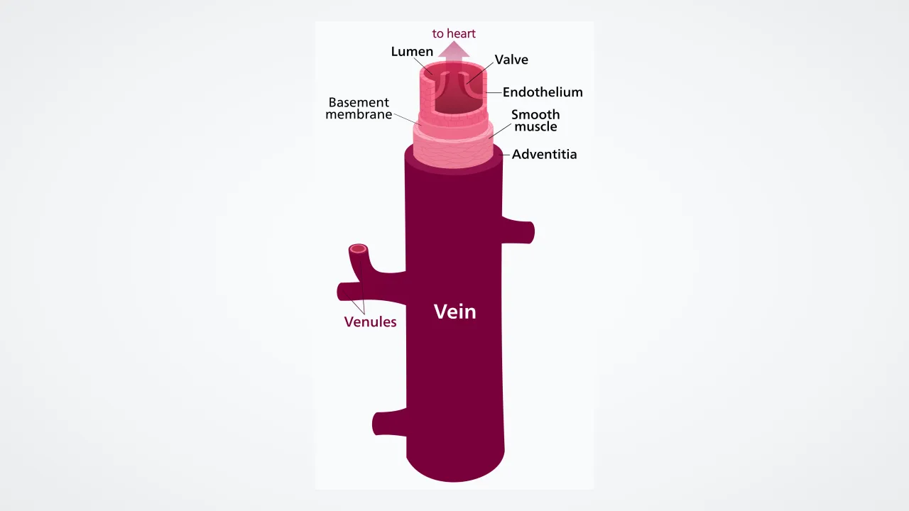
Compared side by side with a matching artery, a vein has a noticeably thinner muscular middle coat and a much wider, collapsible central lumen. Because veins stretch eight times more easily than arteries under small pressure rises, physiologists call them ' vessels.' Storing roughly sixty-five percent of your five-liter blood supply, veins can tighten their smooth muscle walls on command during heavy exercise to mobilize an instant extra liter of blood into active circulation!
2
Twin-Pocket Bicuspid Valves
Inside medium limb veins, folds of the smooth inner tunica intima form pairs of half-moon pockets facing toward the heart. Flowing blood flattens these smoothly against the walls on the way up; if gravity pulls the column downward between muscle steps, blood fills the pockets and seals the vein shut like twin canal gates.
3
The Skeletal Muscle and Respiratory Pumps
Working together with those valves are two clever auxiliary pumps. Deep veins run sandwiched right between bulky calf and thigh muscles, so every stride compresses them to 90 mmHg locally and shoots blood past the next valve rung. Meanwhile, inhaling lowers chest pressure while gently squeezing abdominal veins—sucking venous blood straight into the chest!
4
William Harvey's Forearm Experiment
In 1628, English physician William Harvey pressed two fingers onto a volunteer's forearm vein, slid one finger away from the heart past a valve bump, and watched the vein stay empty until he lifted the valve finger—proving forever that venous blood flows strictly one way toward the heart!
1. Why do physiologists call veins 'capacitance vessels'?
2. What did William Harvey prove with his famous 1628 forearm vein experiment?
🤯 Mind-Blowing Fact • Grade 5
Unlike almost every other vein that heads straight back to the heart, your hepatic portal vein makes a special detour through your liver first so liver cells can inspect and store freshly digested lunch nutrients!
✨ Word Sparks (Grade 5 Glossary)
Tap 🔊 to hear
compliance
ease with which a vessel stretches open
hepatic portal vein
special vein routing gut nutrients through liver
hydrostatic pressure
downward fluid weight caused by Earth's gravity
🪝
How do low-pressure vessels operating at just 10 mmHg return five liters of blood per minute uphill from your ankles?
1
Compliance Mechanics and Venomotor Tone
By the time blood traverses systemic capillary beds and enters postcapillary venules, hydrostatic driving pressure has fallen from 95 mmHg to scarcely 12–15 mmHg—and drops near zero right at the right atrium! To conduct five liters per minute under minimal pressure gradients, veins feature wide lumens and thin tunica media layers rich in distensible collagen-elastin networks, yielding vascular roughly twenty times greater than matching arteries. Sympathetic nerve fibers innervating venous smooth muscle adjust 'venomotor tone,' constricting splanchnic and cutaneous reservoirs during sport to boost cardiac preload.
2
Hydrostatic Columns and Valve Competence
Standing upright creates a gravitational hydrostatic column that would push ankle venous pressure toward 90 mmHg if left uninterrupted. Italian anatomist Hieronymus Fabricius first illustrated venous valves ('little doors') in 1603: endothelial folds reinforced by collagen cores spaced every few centimeters along extremity veins. By breaking one tall column of blood into short, valve-supported segments—driven upward by sole and gastrocnemius muscle contractions—walking keeps average foot venous pressure below 25 mmHg.
3
Superficial, Deep, and Portal Networks
Human veins organize into superficial subcutaneous networks (which also shed body heat and provide safe access for routine blood tests), deep veins accompanying major arteries inside muscular fascia, and specialized portal systems—most famously the , which routes nutrient-rich intestinal blood through a second capillary filter inside the liver before entering the inferior vena cava.
4
Optics of Subcutaneous 'Blue' Veins
Spectroscopy confirms deoxygenated venous blood is dark crimson, never blue: longer red wavelengths (~650 nm) penetrate deeper into dermis to be absorbed by deoxyhemoglobin, while shorter blue wavelengths (~470 nm) scatter back to the observer's retina, tricking cortical color-constancy circuits.
5
From Fabricius to Harvey's Revolution
While Fabricius thought venous valves merely slowed downward blood trickle, his former student at Padua—William Harvey—combined valve geometry with stroke-volume math in De Motu Cordis (1628) to establish closed-loop blood circulation.