2. What creates the 'lub-dub' sound a doctor hears with a stethoscope?
🤯 Mind-Blowing Fact • Grade 2
Over an eighty-year lifetime, your four delicate heart valves open and close roughly three billion times without taking a single break!
✨ Word Sparks (Grade 2 Glossary)
Tap 🔊 to hear
leaflet
one single thin flap of a heart valve
heartstrings
tough tiny cords anchoring middle valve flaps
one-way
moving ahead in only a single direction
🪝
Did you know real 'heartstrings' hold your heart's inner doors in place?
1
Traffic Cops of Your Heart
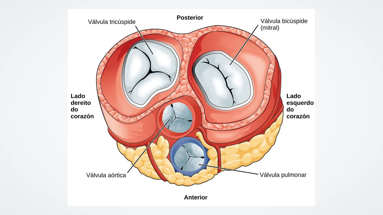
Imagine a busy hallway where everyone must walk in just one direction! Inside your four-roomed heart stand four flexible doors called heart valves. Two sit between the top and bottom rooms, and two guard the big exit tubes.
2
Held by Real Heartstrings
When the strong bottom chambers squeeze, what stops the middle valve flaps from blowing backward like an umbrella in strong wind? Tough little cords—real-life ''—hold the flap edges steady!
3
Moved Only by Blood Push
Amazingly, heart valves have no muscles of their own. Gentle pushes of flowing blood swing them open and click them shut automatically!
1. What job do your tough little 'heartstrings' do inside the heart?
2. What actually pushes your heart valves open and closed?
🤯 Mind-Blowing Fact • Grade 3
Doctor René Laennec invented the first stethoscope in 1816 by rolling up a sheet of paper into a tube so he could hear a patient's heart valves clearly!
✨ Word Sparks (Grade 3 Glossary)
Tap 🔊 to hear
tricuspid
three-flapped valve between right atrium and ventricle
mitral valve
two-flapped valve between left atrium and ventricle
semilunar
exit valve shaped like three half-moon pockets
🪝
Can you name all four one-way doors that keep your bloodstream running on track?
1
Two Doors Between Floors
Picture your heart as a two-story house with a right side and a left side! Between the upper atria and lower ventricles hang two parachute-like doors. On the right stands the valve with three flaps, and on the left stands the with two flaps.
2
Anchored by Chordae Tendineae
When ventricles contract powerfully, tough tendon cords called chordae tendineae—attached to small papillary muscles—hold the and mitral flaps taut. Their firm snap shut creates the first heart sound: 'LUB!'
3
Two Half-Moon Exit Gates
Guarding the exit highways at the top of the ventricles sit two ('half-moon') valves: the pulmonary valve heading to the lungs and the aortic valve heading to the body. Each uses three pocket-shaped cusps.
4
Pocket Catchers Make the 'DUB'
As soon as the ventricles relax, blood tries to slide back down the exit tubes, filling the three half-moon pockets so they seal shut with a crisp 'DUB!'
1. How many flaps do the tricuspid valve and mitral valve each have?
2. Which two valves snap shut to make the second heart sound ('DUB')?
🤯 Mind-Blowing Fact • Grade 4
Although aortic valve leaflets are thinner than a millimeter—roughly the thickness of crisp flower petals—their woven collagen and elastin layers withstand the force of a garden hose blasting seventy times a minute!
Why did a Renaissance anatomist name a left-side heart valve after a bishop's tall pointed hat?
1
Bishop's Hat and Parachute Strings
In the 1500s, pioneering anatomist Andreas Vesalius peered inside the left heart and noticed its two-flapped valve looked remarkably like a bishop's two-pointed ceremonial hat, called a miter—giving the mitral (bicuspid) valve its famous name! Paired with the three-flapped tricuspid valve on the right, these two atrioventricular (AV) valves tether via collagen-rich to muscular pillars called .
2
Why Papillary Muscles Do Not Pull Valves Open
Students sometimes guess that yank AV valves open, yet the exact opposite happens! Valves fall open effortlessly whenever atrial pressure slightly exceeds ventricular pressure. Papillary muscles contract during systole solely to tighten the , preventing up to 120 mmHg of left ventricular pressure from blowing the leaflets backward into the atrium.
3
Three-Pocket Engineering of Semilunar Valves
At the exits to the pulmonary trunk and aorta, the pulmonary and aortic valves need zero strings at all. Built like three-part coat pockets sewn in a ring, their thin cusps flatten smoothly against the artery walls as blood blasts out, then balloon inward and press their three rims tightly together the instant pressure reverses.
4
Listening to Heart Murmurs
Through a stethoscope, smooth laminar blood flow is silent; if a valve narrows slightly or lets a tiny trickle leak back, turbulent eddies create a soft 'whoosh' doctors call a heart —many of which in growing kids are completely harmless 'innocent' murmurs!
1. What is the exact job of papillary muscles when the ventricles squeeze?
2. How do the aortic and pulmonary semilunar valves seal shut without any chordae tendineae?
🤯 Mind-Blowing Fact • Grade 5
Nearly 500 years before modern ultrasound scanners existed, Leonardo da Vinci used glass tubes and floating grass seeds to discover the exact swirling vortices that help close your aortic heart valve!
three aortic pockets creating valve-closing whirlpools
auscultation
medical skill of listening to internal body sounds
🪝
How do avascular leaflets thinner than a dime survive three billion high-pressure flex cycles without fatiguing or cracking?
1
Trilaminar Extracellular Matrix Mechanics
Engineers designing mechanical valves out of titanium and pyrolytic carbon marvel at native human heart valves: living sheets scarcely 0.5 millimeters thick that flex over forty million times every year for nearly a century. Each cusp or leaflet is virtually avascular—nourished directly by oxygen diffusing from passing blood—and laminated into three specialized layers: a load-bearing collagenous facing high back-pressure, a shock-absorbing proteoglycan spongiosa in the center, and an elastin-rich ventricularis (or atrialis) that snaps leaflets smoothly back into shape.
2
Living Interstitial Remodeling
Unlike synthetic plastic flaps that slowly wear down, native valves teem with valvular interstitial cells coated by frictionless endothelial cells. Every day, these resident cells repair microscopic collagen wear caused by cyclic bending stresses—supported by a tough fibrous skeleton of four collagen rings (annuli) that also electrically insulate the upper atria from the lower ventricles.
3
Leonardo da Vinci's Vortex Discovery
How does the aortic valve close so swiftly that almost zero blood leaks backward? Around 1513, Renaissance polymath Leonardo da Vinci built glass models of an ox aorta seeded with grass seeds and sketched swirling eddies—now called Valsalva vortices—forming inside three small bulges () right behind the aortic cusps! Modern MRI confirmed Leonardo was right: spinning vortices gently float the cusps nearly shut even before ventricular ejection ends, and feed oxygen-rich blood directly into the coronary arteries.
4
Auscultation of S1 and S2
Clinically, closure of the mitral and tricuspid valves generates the first heart sound (S1), marking the start of isovolumetric contraction, while closure of the aortic and pulmonic valves produces the crisp second sound (S2) at the onset of diastole.
5
William Harvey and Modern Valve Repair
Studying one-way valves in hearts and veins enabled English physician William Harvey in 1628 to prove blood circulates in a closed loop rather than sloshing back and forth—paving the way for modern catheter-guided valve replacements.