1. What fills the inside of your snail-shaped cochlea?
2. What animal shell gave the cochlea its fun name?
🤯 Mind-Blowing Fact • Grade 2
Birds and fish CAN regrow damaged cochlear hair cells in just a few weeks—and scientists study them to learn how to help human ears heal too!
✨ Word Sparks (Grade 2 Glossary)
Tap 🔊 to hear
pitch
How high or low a sound note feels
bundle
Small neat group standing close together
current
Steady flow of liquid moving forward
🪝
How does a pea-sized spiral sort squeaky flutes from booming drums like a rolled-up piano?
1
Rolled Up Like a Piano
Imagine rolling a whole piano keyboard into two and a half neat spiral turns so it fits inside a tiny pea! Deep inside your skull bone, your snail-shaped cochlea sorts every musical note and spoken word you hear.
2
Waves Bending Tiny Bundles
When the stirrup bone taps the cochlea's front window, waves roll through watery fluid inside. Sixteen thousand microscopic sensor cells topped with tiny 'hair' sway in the like seaweed in ocean surf!
3
Protect Your Hair Cells!
Every time those bend, they send electrical sparks up your hearing nerve. Because humans cannot regrow broken hair cells, keeping music at safe volumes keeps them dancing for life!
1. Why is it super smart to keep headphone volume safe and wear ear protection around roaring engines?
2. About how many spiral turns does a human cochlea make?
🤯 Mind-Blowing Fact • Grade 3
Your cochlea is SO good at its job that in a super-quiet soundproof chamber, a healthy ear can actually hear subtle blood flow and air molecules bouncing inside the ear canal!
✨ Word Sparks (Grade 3 Glossary)
Tap 🔊 to hear
basilar
Flexible inner ribbon sorting high and low pitches
stereocilia
Tiny sensing bristles topping cochlear hair cells
implant
Helpful medical device placed gently inside body
🪝
Where inside your snail-shell cochlea do high violin notes land versus deep thunder rumbles?
1
Hardest Bone Vault in the Body
Knock gently behind your ear and feel your skull's temporal bone—the densest, hardest bone in the human skeleton! Carved right inside that rocky vault sits your fluid-filled cochlea, right next door to three fluid loops that sense your balance.
2
High Notes Near, Low Notes Far
Running down the middle of the snail spiral stretches a flexible ribbon called the membrane. Stiff and narrow near the entrance, it vibrates right away for high-pitched whistles and violins. Wide and floppy up at the spiral's tip, it sways for deep bass guitars and rolling thunder!
3
The Organ of Corti
Perched atop that vibrating ribbon sits the organ of Corti, holding rows of microscopic hair cells. Tiny bristles on top——lean when waves pass, popping open electrical gates!
4
Bionic Cochlear Implants
For kids or adults born with non-working hair cells, doctors can thread a tiny 'cochlear ' wire inside the spiral to spark the auditory nerve directly!
1. Where along the cochlea's spiral ribbon do deep, low-pitched bass notes vibrate best?
2. How does a cochlear implant help someone whose hair cells don't work?
🤯 Mind-Blowing Fact • Grade 4
Newborn hearing screenings take just minutes while a baby sleeps by placing a tiny microphone in the ear to listen for soft 'otoacoustic' echoes produced by dancing outer hair cells!
✨ Word Sparks (Grade 4 Glossary)
Tap 🔊 to hear
tonotopic
Organized neatly step-by-step according to sound pitch
Did you know your cochlea literally MAKES faint sounds of its own while helping you hear?
1
Oval Window In, Round Window Out
Ever try squeezing a sealed plastic bottle filled to the brim with water? Water practically refuses to squish! Because your bony cochlea is completely filled with fluids called perilymph and , every inward push of the stapes at the upper 'oval window' needs a matching pressure relief valve. Right below sits a flexible membrane called the 'round window' that bulges outward in sync, letting fluid waves surge freely down the spiral chambers!
2
Tonotopic Map: One Place Per Pitch
Running thirty-three millimeters along the coil, the basilar membrane widens fivefold from base to apex—acting like an upside-down harp whose short, tight strings sit at the entrance (20,000 Hz) and long, loose strings sit at the tip (20 Hz). Neuroscientists call this neat pitch-to-place layout organization, and your auditory nerve preserves that exact map all the way into your brain's temporal lobe!
3
Inner Listeners Versus Dancing Amplifiers
Inside the organ of Corti, hair cells split into two surprising jobs! One single row of 3,500 'inner hair cells' sends ninety-five percent of all sound messages to your brain. Beside them stand three rows of 12,000 'outer hair cells' that physically dance—lengthening and shortening thousands of times per second like tiny motors to amplify quiet whispers a hundredfold!
4
Otoacoustic Emissions: Ears That Hum Back!
Because outer hair cells actively pump energy, sensitive hospital microphones tucked in a newborn baby's ear can actually record faint echoes bouncing BACK out—painlessly confirming healthy hearing on day one!
1. Why does the fluid-filled cochlea need a flexible 'round window' below the oval window?
2. What surprising mechanical superpower do the three rows of 'outer hair cells' perform?
🤯 Mind-Blowing Fact • Grade 5
Why is your sense of hearing ten times faster at catching split-second timing than your sense of sight? Because nanometer 'tip-link' threads yank cochlear ion gates open mechanically in just TEN MICROSECONDS—no slow chemical chain reaction required!
✨ Word Sparks (Grade 5 Glossary)
Tap 🔊 to hear
mechanotransduction
Turning tiny physical movement straight into electricity
perilymph
Outer fluid carrying pressure waves through cochlea
prestin
Ultrafast motor protein powering outer hair amplification
🪝
How do nanometer molecular 'tip-link' springs and bulletproof prestin motor proteins make your inner ear nature's fastest mechanical sensor?
1
Georg von Békésy and the Traveling Wave
In 1561, Italian anatomist Gabriele Falloppio named the coiled bony labyrinth 'cochlea' after a land snail, and three centuries later Alfonso Corti mapped its delicate cellular ribbon under a microscope. Yet how pitch separated inside remained mysterious until Hungarian biophysicist Georg von Békésy—awarded the 1961 Nobel Prize—strobed microscopic silver flakes along the basilar membrane! Békésy watched each frequency launch an asymmetric 'traveling wave' cresting at one exact tonotopic coordinate before dying abruptly beyond it.
2
Three Fluid Chambers and the Endocochlear Battery
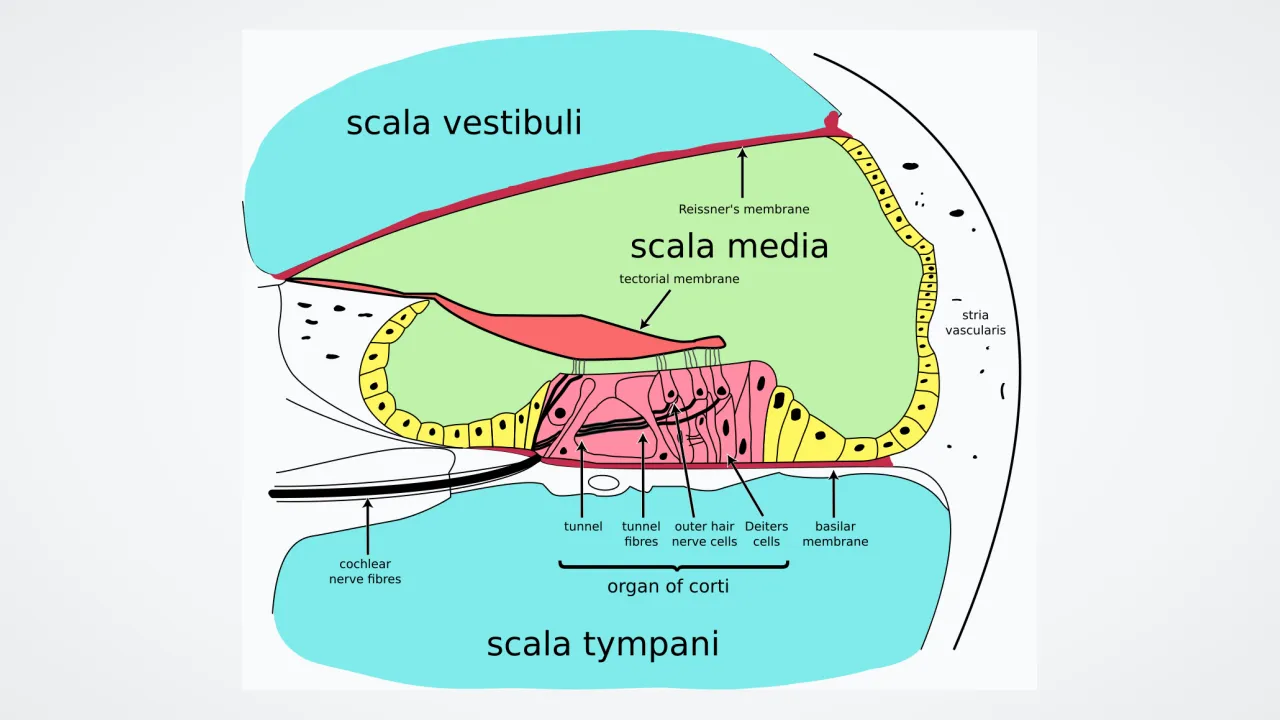
Cross-sectioning the spiral reveals three parallel fluid ducts: scala vestibuli and scala tympani filled with sodium-rich , sandwiching the central scala media filled with potassium-rich endolymph. Along the outer wall, a vascular powerhouse called the stria vascularis pumps potassium ions into endolymph to maintain a +80-millivolt 'endocochlear potential'—the highest extracellular voltage anywhere in the human body!
3
Tip Links: Mechanotransduction in Microseconds
Unlike sight or smell—which rely on multi-step chemical cascades taking milliseconds—hearing uses direct mechanical spring gates! Stepping upward like organ pipes atop each hair cell, stereocilia connect to taller neighbors via five-nanometer protein threads called tip links. When shearing fluid against the tectorial membrane leans a bundle by just nanometers, tip links yank ion channels physically open in ten microseconds—ten times faster than photoreceptors—releasing glutamate onto spiral ganglion neurons of cranial nerve VIII!
4
Prestin Motors and the Cochlear Amplifier
In 1978, physicist Thomas Gold's bold prediction of an active ear amplifier was vindicated when David Kemp recorded otoacoustic emissions! Packed into outer hair cell walls is a unique piezoelectric-like motor protein appropriately named '' (from musical 'presto' for fast!), which changes length at kilohertz speeds without burning ATP—amplifying faint whispers by forty decibels (100× amplitude) and sharpening pitch selectivity so musicians distinguish notes fractionally apart.
5
Neuroprosthetics and Gene Therapy Horizons
Modern multi-channel cochlear implants—pioneered by Graeme Clark, Ingeborg Hochmair, and Blake Wilson—exploit tonotopy by routing filtered frequency bands to targeted electrodes along scala tympani, restoring speech to over a million recipients worldwide while researchers test ATOH1 and OTOF gene therapies to regenerate natural hair-cell synapses!