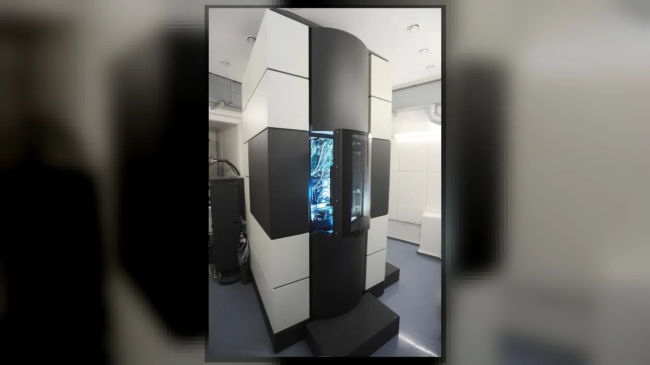
1. Why cannot an ordinary glass light microscope see the tiniest viruses?
2. What steers and focuses the beam of electrons inside the tall metal column?
🤯 Mind-Blowing Fact • Grade 2
Every picture taken by an electron microscope starts out completely black and white because electrons are smaller than colors of light—scientists add color later on computers!
✨ Word Sparks (Grade 2 Glossary)
Tap 🔊 to hear
vacuum
An empty space with all air pumped out
microbe
A living thing too small to see alone
beam
A focused stream of particles or light
🪝
If you magnified an ant until it looked as long as a school bus, what secret textures would pop out?
1
Hitting the Limit of Light
Peer at pond water under a school microscope, and swimming wiggle into view! Yet even the sharpest glass lens blurs out around two thousand times magnification because light waves are too chunky for smaller details.
2
Swapping Light for Electrons
In 1931, German engineers Ernst Ruska and Max Knoll tried a bold new . Electrons travel in waves thousands of times narrower than visible light, letting scientists zoom past one million times!
3
Pumping Out All the Air
Inside the tall steel tower, powerful pumps suck out all the air to create a so electrons will not bump into air molecules on their trip to the target.
1. Why are all the air molecules pumped out of an electron microscope's column?
2. Why are raw electron microscope photos always in shades of gray instead of natural color?
🤯 Mind-Blowing Fact • Grade 3
To photograph a fuzzy pollen grain or spider fang in 3D without electric static building up, scientists often mist it with a layer of pure gold only a few atoms thick!
✨ Word Sparks (Grade 3 Glossary)
Tap 🔊 to hear
wavelength
Distance from one wave crest to next
electromagnet
Wire coil that becomes magnetic with electricity
virus
Tiny infectious agent smaller than a cell
🪝
Why could no scientist on Earth see what a virus actually looked like until the 1930s?
1
Trying to Feel Pebbles With Boxing Gloves
Imagine wearing giant puffy boxing gloves and trying to feel tiny Braille dots on an elevator button! That was the exact problem biologists faced with light microscopes: waves of visible light were simply too wide to trace tiny or parts inside cells.
2
Ruska and Knoll's Magnetic Lenses
Physicists discovered in the 1920s that fast-moving electrons travel like ultra-short waves—up to 100,000 times shorter than light! In 1931 in Berlin, young engineer Ernst Ruska and Max Knoll wound copper coils into ring-shaped that focused an electron beam just like glass lenses focus sunlight.
3
Two Ways to Look Close
Scientists soon developed two main styles. A Transmission Electron Microscope (TEM) shoots electrons straight through paper-thin slices to reveal inner cell parts and atoms. A Scanning Electron Microscope (SEM) sweeps a beam across gold-dusted surfaces, creating jaw-dropping 3D views of compound insect eyes, pollen spikes, and computer chips!
4
Seeing Invisible Invaders
Within years, Ernst Ruska's brother Helmut captured the very first photographs of , transforming medicine forever.
1. Which type of electron microscope sweeps across an object's outside surface to build dramatic 3D-looking pictures?
2. Why do technicians coat bugs or pollen in an ultra-thin mist of gold before scanning them?
🤯 Mind-Blowing Fact • Grade 4
Special diamond knives—sharper than steel scalpels—are used to slice biological samples nearly a thousand times thinner than a human hair so electrons can zip right through!
✨ Word Sparks (Grade 4 Glossary)
Tap 🔊 to hear
nanometer
One billionth of a single meter
diffraction
Bending and blurring of waves around tiny edges
filament
Thin heated wire that releases stream of electrons
🪝
How did quantum physics turn negatively charged particles into a microscope five thousand times sharper than glass?
1
Abbe's Unbreakable Wall of Light
In 1873, German physicist Ernst Abbe proved a startling mathematical law: no optical microscope, no matter how flawlessly polished its glass lenses, could ever separate two dots closer than roughly 200 —half the wavelength of violet light. Millions of life's tiniest structures lay hidden behind that wall.
2
Wave-Particle Duality Opens the Door
Fifty years later, Louis de Broglie proposed that moving matter particles—including electrons—also behave like waves, and the faster they accelerate, the shorter their wavelength shrinks! In 1931 at Berlin's Technical University, Ernst Ruska and Max Knoll accelerated electrons from a heated tungsten through high voltage and focused them with doughnut-shaped magnetic coils, building the first prototype electron microscope. By 1933, Ruska surpassed the optical resolution limit forever.
3
Inside TEM and SEM Columns
Accelerated by 100,000 to 300,000 volts inside an ultra-high vacuum, electrons shrink to wavelengths smaller than five trillionths of a meter! In a Transmission Electron Microscope, the beam pierces diamond-sliced specimens thinner than 100 to map crystal lattices and ribosomes. In a Scanning Electron Microscope, raster coils steer a needle-thin beam back and forth while detectors count secondary electrons knocked from the surface.
4
Flash-Freezing Life in Action
Modern cryo-electron microscopy flash-freezes delicate proteins in glassy water at minus 196 degrees Celsius, letting researchers map vaccine targets atom by atom.
1. What happens to the wavelength of an electron beam when scientists crank up the accelerating voltage?
2. How does modern cryo-electron microscopy keep delicate watery proteins from shriveling inside the vacuum?
🤯 Mind-Blowing Fact • Grade 5
With modern magnetic aberration correctors—acting like high-tech eyeglasses for electron beams—microscopes can measure distances between atoms to within half the width of a single hydrogen atom!
✨ Word Sparks (Grade 5 Glossary)
Tap 🔊 to hear
picometer
One trillionth of a meter in length
aberration
Lens imperfection that slightly blurs sharp focus
vitrify
Freeze water into smooth crystal-free glass
🪝
Why does accelerating an electron to seventy percent of the speed of light allow scientists to photograph columns of individual atoms?
1
Shattering the Optical Diffraction Barrier
Every imaging instrument is governed by wave diffraction: Ernst Abbe demonstrated in 1873 that resolving power cannot exceed roughly half the illuminating wavelength (lambda / 2NA). Because visible photons span 400 to 700 nanometers, optical microscopes blur anything finer than ~200 nanometers—leaving viruses, lipid bilayers, semiconductor gates, and crystal defects completely invisible.
2
De Broglie Waves and Magnetic Lenses
French physicist Louis de Broglie unlocked the escape route in 1924 by proving that every particle possesses a wavelength inversely proportional to its momentum (lambda = h/p). After Hans Busch showed in 1926 that axisymmetric magnetic fields refract electron trajectories mathematically like convex glass lenses refract light, Berlin engineers Max Knoll and Ernst Ruska constructed the first two-lens transmission electron microscope in April 1931—work that earned Ruska the 1986 Nobel Prize in Physics.
3
Relativistic Beams in Ultra-High Vacuum
At the crown of a modern microscope, a Schottky field-emission gun extracts a coherent beam of electrons and accelerates them across 200,000 volts to roughly 70 percent of light speed, compressing their wavelength to 2.5 —nearly forty times smaller than a hydrogen atom! Pumped to $10^{-7}$ pascals so electrons travel unimpeded by gas molecules, the column uses electromagnetic condenser, objective, and projector coils—paired with computer-controlled multipole correctors—to focus the beam within 0.05 nanometers.
4
TEM, SEM, and Cryo-EM Revolutions
Ultramicrotome diamond blades shave specimens under 100 nanometers thick for Transmission Electron Microscopy (TEM) and Scanning Transmission modes that map atomic columns and elemental X-ray signatures. Meanwhile, Scanning Electron Microscopes (SEM) raster lower-energy beams across conductive surfaces to detect secondary electrons for three-dimensional topography, and Nobel-winning Cryo-EM vitrifies hydrated biomolecules in liquid ethane (-188 °C) to reconstruct 3D protein machinery.
5
Engineering the Nano-Age
From inspecting 2-nanometer smartphone transistors and lithium-battery interfaces to mapping viral spike proteins within days, electron optics underpins modern nanotechnology, materials science, and structural biology.