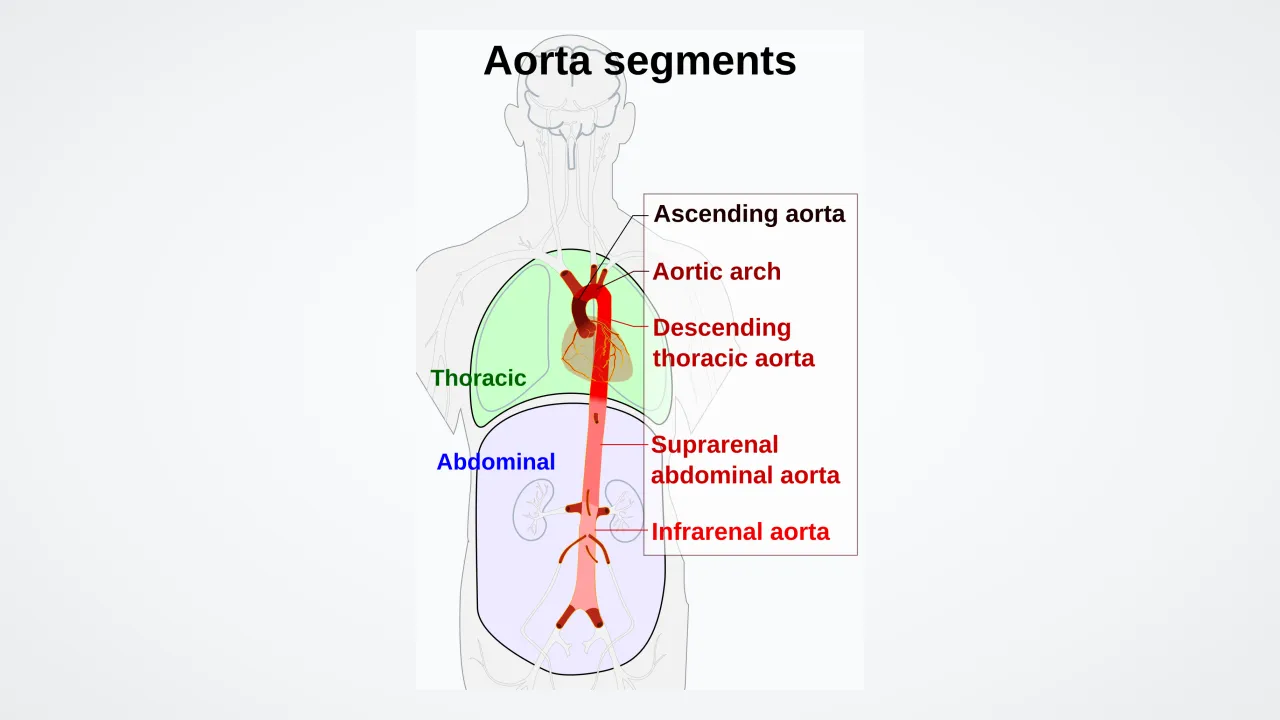
1. What shape does the top of your aorta make above your heart?
2. What kind of blood rushes through your aorta?
🤯 Mind-Blowing Fact • Grade 2
Over an average human lifetime, your aorta carries enough blood to fill an entire supertanker ship!
✨ Word Sparks (Grade 2 Glossary)
Tap 🔊 to hear
artery
blood vessel carrying blood away from heart
elastic
able to stretch wide and bounce back
trunk
main central stem before smaller branches split
🪝
Which body part gets the very first sip of fresh blood from your giant aorta?
1
Superhighway Number One
Feel your heart thump after a race! Your strong left ventricle launches fresh, oxygen-packed blood straight into your body's largest , the aorta. Think of it as the main of a giant tree before all the branches split off.
2
Feeding the Heart First
Guess who gets the very first tiny side branches off the aorta? Your hardworking heart muscle itself! Next, three big top branches send blood rushing upward to your brain and both arms.
3
Stretchy and Bouncy Walls
Your aorta has thick, rubbery walls. It stretches wide when blood blasts in, then squeezes back gently to keep blood gliding forward between beats!
1. Which organ receives the very first tiny branches right at the base of the aorta?
2. Why does the aorta need super stretchy, elastic walls?
🤯 Mind-Blowing Fact • Grade 3
A blue whale's aorta is over nine inches (23 centimeters) wide—wide enough for a small child to crawl through like a playground tunnel!
✨ Word Sparks (Grade 3 Glossary)
Tap 🔊 to hear
ascending
climbing or rising upward from the bottom
thoracic
relating to the chest area inside ribs
diaphragm
dome-shaped breathing muscle beneath your lungs
🪝
How does one giant blood vessel supply your brain, stomach, kidneys, and toes all at once?
1
Four Named Highway Zones
Trace an imaginary line from the middle of your chest up toward your collarbone and straight down to your bellybutton! You just mapped the route of your aorta. Anatomists name its four main stretches along the way: the aorta, aortic arch, aorta, and abdominal aorta.
2
Upstairs Exits From the Arch
Right after feeding the heart via coronary arteries, the curving aortic arch sends three major highways upward—delivering high-priority oxygen to your hungry brain, neck, shoulders, and hands.
3
Down Through the Diaphragm
Diving downward behind your heart, the aorta slips through a special opening in your breathing muscle—the —becoming the abdominal aorta. Side exits feed your liver, stomach, intestines, and kidneys before splitting in two near your bellybutton for both legs!
4
Built-In Pressure Sensors
Special nerve sensors in the aortic arch constantly measure blood pressure and alert your brainstem in a flash if you stand up fast.
If your heart stops squeezing between beats, why doesn't blood stop moving inside your arteries?
1
The Elastic Windkessel Effect
Watch an old-fashioned fire engine pump water: without an air-cushioned chamber, water would shoot out in jerky spurts instead of a smooth stream! German physiologists named your aorta's matching superpower the Windkessel ('air chamber') effect. When your left ventricle blasts seventy milliliters of blood into the aorta during systole, the aorta balloons outward, storing nearly half that stroke's energy in its stretchy walls.
2
Fifty Layers of Elastin Springs
Then, the instant the aortic valve snaps shut for diastole, the stretched aortic wall recoils inward like a giant elastic sleeve! That smooth squeeze keeps blood gliding continuously forward into your brain and kidneys even while your heart rests between beats. Under a microscope, the aorta's middle wall (tunica media) packs forty to seventy concentric sheets of elastin fibers interleaved with smooth muscle and tough collagen.
3
Vessels That Feed a Vessel
Because the adult aortic wall is roughly two millimeters thick, outer wall cells sit too far from rushing blood inside to get oxygen by simple diffusion. Instead, tiny microscopic blood vessels called ('vessels of the vessels') weave through the outer aortic coat to keep the superhighway itself nourished!
4
Baroreceptor Reflexes Against Dizziness
Embedded inside the aortic arch wall are stretch-sensitive . Stand up abruptly from bed, and if upper-body pressure dips even slightly, aortic baroreceptors signal your brainstem via the vagus nerve to quicken your pulse so you never black out.
1. How does the aorta's Windkessel effect protect your brain and kidneys?
2. Why does the thick outer wall of the aorta need tiny 'vasa vasorum' vessels?
🤯 Mind-Blowing Fact • Grade 5
Most of the springy elastin protein molecules inside your aorta were built before you finished growing—and those exact same molecules keep stretching and snapping back over two billion times across your lifetime!
✨ Word Sparks (Grade 5 Glossary)
Tap 🔊 to hear
tunica media
thick middle artery layer packed with elastin
chemoreceptor
sensor monitoring blood oxygen and carbon dioxide
lamellae
thin concentric sheets of elastic tissue
🪝
How do sixty concentric sheets of rubber-like protein convert violent 120 mmHg heart surges into gentle capillary flow?
1
Trilaminar Architecture of an Elastic Artery
Classified histologically as the body's premier conducting (elastic) artery, the human aorta spans roughly thirty centimeters in length and up to three centimeters in lumen diameter at its root. Its wall organizes into three tunics: a slick endothelial tunica intima minimizing shear drag, a thick composed of forty to seventy fenestrated concentric of elastin alternating with vascular smooth muscle and Type I/III collagen, and an outer collagenous tunica adventitia nourished by vasa vasorum.
2
Biomechanics of Elastin vs. Collagen Recruitment
Why doesn't the aorta balloon endlessly and burst during heavy sprinting? Its two structural proteins work as a two-stage safety harness! At normal physiological pressures (80 to 120 mmHg), extensible elastin bear the load, expanding easily to absorb left ventricular stroke energy. If blood pressure surges higher, stiff wavy collagen jackets straighten out and lock tight, preventing dangerous overexpansion while diastolic recoil propels blood into the coronary arteries and systemic tree.
3
Anatomical Landmarks and Chemoreceptors
Rising from the aortic annulus, the ascending aorta gives off the right and left coronary arteries from the sinuses of Valsalva before curving posteriorly as the aortic arch—dispatching the brachiocephalic trunk, left common carotid, and left subclavian arteries. Nestled beneath the arch sit tiny aortic bodies containing that monitor arterial oxygen, carbon dioxide, and pH alongside stretch-sensing baroreceptors.
4
Arterial Stiffening Across Lifespans
Because elastin fibers are synthesized almost entirely before adulthood with a half-life exceeding seventy years, decades of billions of stretches gradually fray elastin, explaining why pulse pressure widens in elderly grandparents.
5
From Aristotle's Naming to Otto Frank's Math
Greek philosopher Aristotle first coined the word 'aorta' (meaning 'to lift or suspend,' as the heart seemed hung from it) in the fourth century BCE; English clergyman-scientist Stephen Hales measured arterial Windkessel cushioning in 1733, and German physiologist Otto Frank formulated its exact mathematical equations in 1899.