🧬 HUMAN BODY
🧬 Pulmonary Alveoli • Respiratory System
VA Science & Health SOL 1.1, 3.1, 4.2, 5.1 (Cells, Organs & Body Systems)
Alveoli Air Sacs
Half a Billion Microscopic Grape Clusters Trading Every Breath
Grade 4 • Expanding • 195 words
🩺 Body System & LocationRespiratory System • Clustered at the very tips of bronchioles inside both lungs
🔬 Size / Cell Count300–500 million air sacs • ~750 sq ft (70 sq m) combined surface area
⚙️ Core JobSwaps fresh inhaled oxygen into blood capillaries while trading out carbon dioxide
⚡ Body SuperpowerPacks the surface area of half a tennis court tightly inside two spongy chest organs
🤯 Mind-Blowing Fact • Grade 1
Your two lungs hold hundreds of millions of tiny alveoli air sacs—that is why lungs feel soft like sponges!
✨ Word Sparks (Grade 1 Glossary)
Tap 🔊 to hear
Alveoli
Tiny bubble-like air pockets inside your lungs
Swap
To trade one thing for another thing
Pocket
A small hollow pouch that holds something
🪝
Why do your lungs feel soft and spongy instead of hollow like two big balloons?
1
Tiny Air Bubbles
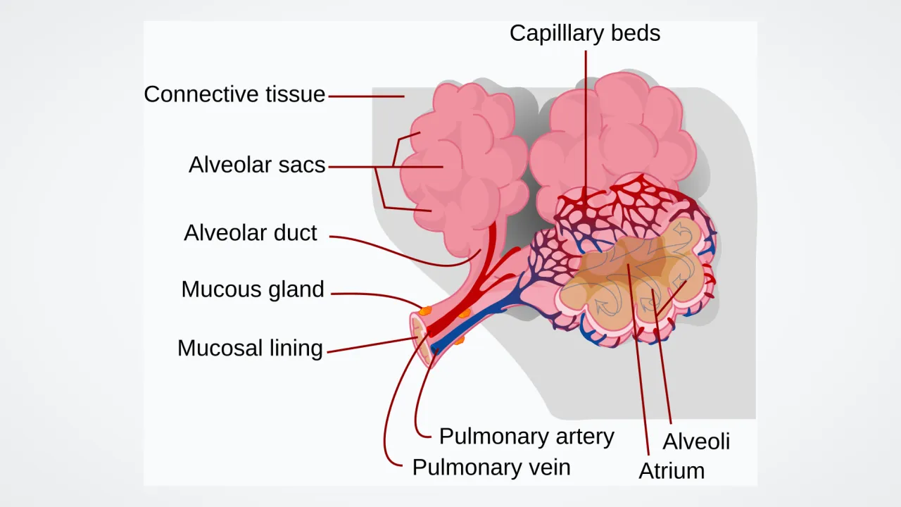
Zoom all the way to the tips of your lung twigs! Waiting there are millions of tiny round called . They look just like bunches of tiny bubbles or grapes.
2
The Great Air Swap
Thin blood tubes hug every single bubble. Fresh oxygen steps out of the bubble into your blood, and used air steps into the bubble to leave!
3
Puffing In and Out
Every time you breathe in, millions of puff up gently, then let used air whoosh right out.
1. What fruit bunch do clusters of alveoli air sacs look like under a microscope?
2. What important trade happens inside your alveoli air sacs?
🤯 Mind-Blowing Fact • Grade 2
Unfolded flat, all the tiny alveoli inside adult lungs would cover roughly half a tennis court!
✨ Word Sparks (Grade 2 Glossary)
Tap 🔊 to hear
Capillary
The tiniest, thinnest blood tube in your body
Surface area
Total flat space covering the outside or inside
Exhale
To breathe used air gently out of lungs
🪝
How could something as big as a tennis court fit neatly inside your chest?
1
Millions of Mini Rooms
If your lungs were just two empty balloons, they would not have nearly enough wall space to share oxygen fast enough! Instead, your lungs are packed with 300 to 500 million microscopic air sacs called alveoli. Flattened out side by side, their walls would cover half a tennis court!
2
Walls Thinner Than Tissue Paper
Wrapped around every air sac is a fine net of tiny blood tubes called capillaries. Both walls are fifty times thinner than a sheet of tissue paper, so gases slip across in a blink!
3
Trading Places
Oxygen glides from the air sac into passing red blood cells, while waste carbon dioxide hops into the sac to ride your next outside.
1. Why is it better for lungs to have millions of tiny alveoli instead of being two giant hollow balloons?
2. Why are the walls of alveoli and their surrounding capillaries ultra-thin?
🤯 Mind-Blowing Fact • Grade 3
Special lung cells make a natural soapy coating called surfactant so half a billion moist air sacs pop open effortlessly on every breath!
✨ Word Sparks (Grade 3 Glossary)
Tap 🔊 to hear
Alveolus
Singular word for just one tiny air sac
Diffusion
Natural spreading of gas from crowded to uncrowded
Surfactant
Soapy lung coating keeping wet air sacs open
🪝
Why don't wet microscopic air bubbles inside your lungs stick shut like a wet plastic bag?
1
End of the Breathing Trail
Follow a molecule of oxygen all the way down your windpipe, through branching bronchi, and out the tip of a hair-thin bronchiole. Welcome to an —one of nearly half a billion microscopic air sacs where respiratory magic happens!
2
Diffusion Without Pumps
No tiny fans or motors push oxygen into your blood. Instead, alveoli use natural : molecules naturally spread from crowded areas to less crowded areas. Freshly inhaled air in an is packed with oxygen, so oxygen glides across the ultra-thin barrier into oxygen-hungry red blood cells, while crowded carbon dioxide in the blood diffuses out into the sac.
3
The Non-Stick Soap Secret
Have you ever tried pulling apart a wet plastic sandwich bag? Water droplets cling tightly together! To keep moist alveoli from sticking shut when you exhale, special cells coat every sac with a soapy natural liquid called .
4
Elastic Springs
Webs of stretchy elastic fibers wrap outside each cluster, helping alveoli spring gently back into shape with every breath.
1. How does oxygen move from an alveolus into a red blood cell?
2. What would happen inside moist alveoli without their soapy surfactant coating?
🤯 Mind-Blowing Fact • Grade 4
Even when you sprint at top speed and blood zips past an alveolus in just 0.25 seconds, the barrier is so thin that red cells still fill up completely with oxygen!
✨ Word Sparks (Grade 4 Glossary)
Tap 🔊 to hear
Pneumocyte
Specialized lung cell building or coating an alveolus
Surface tension
Clinging pull between water molecules on wet surfaces
Barrier
A thin dividing wall between two neighboring spaces
🪝
How can a red blood cell load up completely on fresh oxygen during a pit stop lasting just a quarter of a second?
1
An Ultra-Thin Crossing Bridge
At rest, a red blood cell glides through the capillary mesh wrapping an alveolus in about 0.75 seconds—and during hard sprinting, that pit stop shrinks to just 0.25 seconds! Yet hemoglobin still leaves nearly 100 percent loaded with oxygen. Why? Because the blood-air separating alveolar air from blood plasma is barely half a micrometer thick—roughly one-hundredth the width of a human hair!
2
Three Specialist Cell Neighbors
Zooming into a single alveolus reveals three remarkable cell partners sharing the neighborhood. Paper-flat Type I form 95 percent of the sac's wall, making the diffusion bridge ultra-thin. Tucked into corners, cube-shaped Type II pneumocytes manufacture pulmonary surfactant—a phospholipid-protein mix that breaks water so smaller alveoli do not collapse into larger ones. Type II cells can even divide to replace injured Type I cells!
3
Dust-Busting Alveolar Macrophages
Because cilia stop up in the bronchioles, who cleans the alveoli themselves? Wandering white blood cells called alveolar macrophages ('dust cells') patrol inside the air sacs, gobbling up any stray microscopic specks or bacteria that made it all the way down.
4
Shortcut Windows Between Sacs
Tiny side openings called pores of Kohn even link neighboring alveoli together, equalizing air pressure and providing backup detours if one tiny twig gets blocked!
1. How do Type I and Type II pneumocytes divide up jobs inside an alveolus?
2. Who cleans stray microscopic dust specks out of alveoli where there are no cilia?
🤯 Mind-Blowing Fact • Grade 5
Pediatrician Dr. Mary Ellen Avery's 1959 discovery that premature babies lacked alveolar surfactant launched a simple treatment that has saved millions of tiny lives worldwide!
✨ Word Sparks (Grade 5 Glossary)
Tap 🔊 to hear
Partial pressure
Individual pushing force of one gas in air
Phospholipid
Fat-like molecule forming cell membranes and lung surfactant
Perfusion
Flow of blood through an organ's capillary network
🪝
How did studying soap bubbles and Laplace's physics equation save the lives of millions of premature newborn babies?
1
Fick's Law Perfected in Living Tissue
According to Fick's law of diffusion, gas transfer rate increases directly with surface area and partial-pressure difference while rising inversely with membrane thickness. Human pulmonary alveoli optimize every variable of that equation! Roughly 480 million polyhedral alveoli pack 70 to 100 square meters of gas-exchange area into thoracic lungs, while fusing the Type I alveolar epithelial basement membrane directly to the pulmonary capillary endothelial basement membrane—trimming the respiratory membrane to just 0.2–0.5 micrometers.
2
Partial Pressures Driving Gas Exchange
Within alveolar air, oxygen (PO2) hovers near 104 mmHg compared to 40 mmHg in incoming deoxygenated pulmonary arterial blood, driving rapid passive O2 diffusion into erythrocytes. Although carbon dioxide's pressure gradient is smaller (45 mmHg in venous blood versus 40 mmHg in alveolar gas), CO2 is twenty times more soluble in water than O2, allowing equal volumes of CO2 to exit effortlessly with every breath.
3
Laplace's Law and Mary Ellen Avery's Discovery
By Laplace's law for spheres (P = 2T/r), if surface tension (T) stayed constant, tiny alveoli with smaller radii (r) would generate higher inward collapsing pressure than larger alveoli and snap shut! Dipalmitoylphosphatidylcholine (DPPC) surfactant from Type II pneumocytes solves this by packing tighter as an alveolus shrinks, slashing surface tension near zero. In 1959, pediatrician Dr. Mary Ellen Avery discovered that premature infants struggled to breathe because immature lungs had not yet produced surfactant—leading to modern surfactant replacement therapy that has saved millions of newborns.
4
Ventilation-Perfusion Matching
Alveolar arterioles even auto-tune blood flow: if one cluster of alveoli receives less fresh air, local vessels constrict slightly to redirect blood toward better-ventilated sacs, maximizing oxygenation efficiency.
5
Malpighi's Microscopic Breakthrough
Until Marcello Malpighi examined frog lungs under early microscopes in 1661 and discovered alveoli and capillaries, physicians believed blood mixed openly with air inside spongy lung flesh!